PREVIOUS   NEXT   ISSUE   CONTENTS   HOME 

4.5 Diagenesis and pathologies

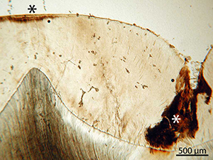
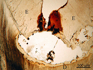
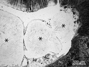
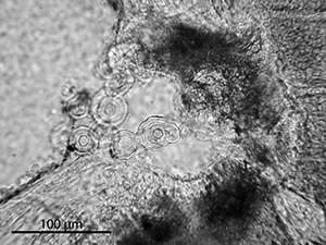
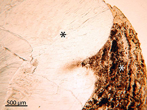
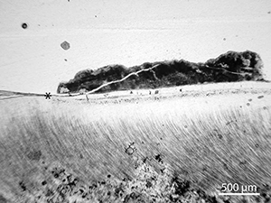
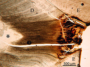
Several types of pathologies were observed and are illustrated here, to allow readers to identify this as non-diagenetic. It is not possible to establish, however, whether superficial etching of the enamel surface is pathology, or diagenetic in nature.

Figure 49
Figure 49: Micrograph of sample EIN-04. In this tooth the crown enamel has been worn down to the dentine during life and the exposed dentine is affected by orange staining and generalised destruction (white asterisk). This is at least partially pathological, seen by the tertiary, or reparative, dentine (black asterisk). This will form as reaction to stimuli such as caries or attrition and will be produced only by the cells affected by the stimulus (Nanci 2003)

Figure 50
Figure 50: Micrograph of sample EIN-17. The white asterisk marks an enamel caries lesion, clearly identifiable with a reaction (orange staining) in the dentine underneath (Marsland and Browne 1975). It is not possible to determine whether or not the enamel surface etching and staining (black asterisk) is a smooth surface lesion, or diagenetic in origin

Figure 51
Figure 51: Micrograph of sample EIN-20. An extensive lesion of the dentine has undermined the enamel adjacent to the fissure. Such lesions may cause demineralisation of a large area directly underneath it (Marsland and Browne 1975). The lesion contains inorganic diagenetic inclusions

Figure 52
Figure 52: Micrograph of sample EIN-20. Three large pulp stones (asterisks) within the pulp cavity. These may arise as an age change or accompany inflammatory or degenerative changes in the pulp. Calcification of the pulp results in the formation of discrete, approximately circular mineralised masses (Marsland and Browne 1975). Finally, these may be completely incorporated into the dentine, as seen in Figure 21

Figure 53
Figure 53: Micrograph of sample SAI-04. A cluster of small pulp stones within the pulp cavity

Figure 54
Figure 54: Micrograph of sample EIN-10. A thick layer of calculus (white asterisk) overlies the enamel (black asterisk)

Figure 55
Figure 55: Micrograph of sample EIN-12. Calculus overlying the cementum just below the enamel-cementum junction (asterisk)