
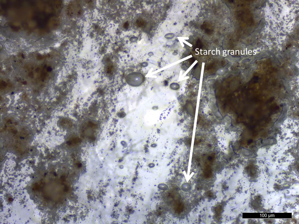
Each experimental flake was handled with care to prevent contamination from hands and cross-contamination between samples. This enabled us to limit the introduction of skin flakes from bare hands, which Pedergnana et al. (2016) recently identified as common contaminants on stone tools. Likewise, we did not use modelling clay. Blu-Tack® was instead used as the base support material during microscopic examination of the experimental flakes. We know that Blu-Tack®; can transfer to the flint, and can also stick to and remove residues from the stone surface. To prevent contamination or loss of residues, the microscope stage was prepared by placing a bed of Blu-Tack®; on the stage and then overlaying a new layer of plastic paraffin film (Parafilm M®) to create a fresh unused surface that separated the mouldable Blu-Tack®; from each flake. Parafilm M® was chosen instead of cling film to place on top of the Blu-Tack®; for the mounting surface because it is flexible and able to withstand stress without tearing or puncturing. Thus, the experimental flakes never came into contact with the Blu-Tack®. The paper-covered side of the parafilm was removed and placed facing upward to be in contact with the specimen and the parafilm was orientated by touching the edges only. Each flake was placed on the microscope stage and manoeuvred using a new powder-free glove each time. These procedures helped to minimise, but did not eliminate, the presence of modern contaminants. For example, blue and pink fibres from clothing were sometimes observed, and starch granules within the glue of the double-sided tape used to mount soil samples on slides were found (Figures 9 and 10).
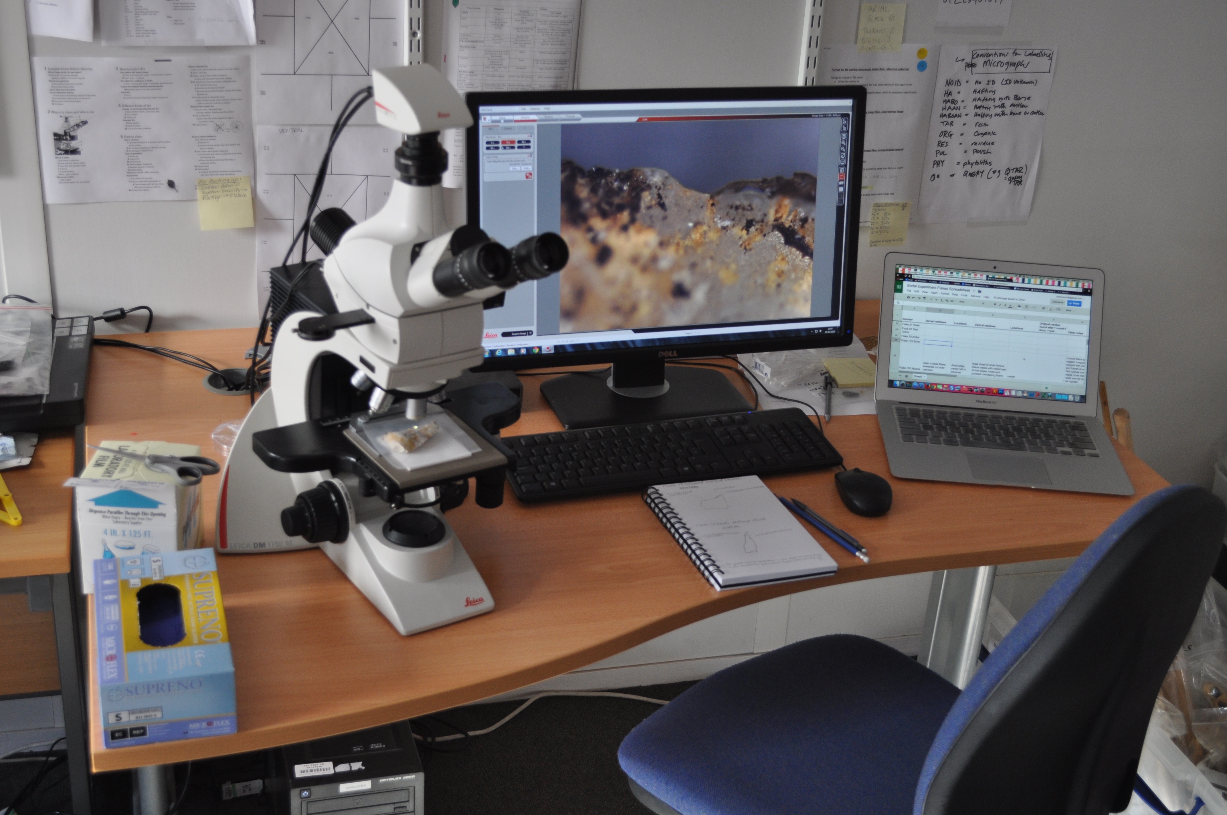
Microscopic analysis was carried out on each buried flake from each unit and time interval. All edges as well as the dorsal and ventral surfaces of the flakes were scanned for residues using a reflected visible light microscope, a Leica DM1750 M, using the 10x and 20x objectives (Figure 11). Microscopic visual characteristics recorded for each residue included colour, shape, texture, brightness, reflectivity, transparency, structural patterns, presence of identifiable cell margins (where possible), presence of microcrystals and their colour and shape, nature of residue deposit edges (circular, ragged, angular, etc.). Langejans 'preservation index' (2010, 978) was used as a guide to rate the state of residue preservation. This index is rated on a scale of 5 (observed immediately after tool use) to 0 (no residues observed), and is subjective, but attempts to quantify the amount of residue left on each flake. A series of z-stacked images was taken for each residue image with the Leica Montage software program. This program stitches together micrographs taken at different planes of focus in the z axis, resulting in an in-focus composite image capturing residue microtopography in full (Monnier et al. 2012). All VLM images presented in this paper are composites.
To assess the quality of residue characterisation attainable, all reference collection residues were then imaged with a variable pressure Hitachi TM-1000 tabletop SEM at LacCore Facility, University of Minnesota (Figure 12). Each residue was imaged in the SEM at multiple magnifications up to 5000x, resulting in a total of 140 separate SEM micrographs. This enabled comparisons between VLM and SEM micrographs as a basis for determining whether SEM analysis shows additional residue features not visible with VLM. Monnier et al. (2012) attempted to answer this same question in their Test 2. Test 2 was a blind test and involved three archaeology graduate students and one archaeology professor, all with no previous experience with residue analysis. Each person was trained by examining reference collection residue micrographs for two individual two-hour sessions prior to the test. The test subjects were presented with a combined set of VLM and SEM images to determine whether the SEM images could improve identification success rates of ambiguous residues over using VLM images alone. SEM images were shown to improve the identifiability of ambiguous residues in the categories of hardwood (Fraxinus sp.), softwood (Picea sp.), and antler, but did not improve the ability of test subjects to identify ambiguous hide and bone residues correctly. Borel et al. (2014) concluded that optical (reflected) VLM and SEM micrographs should be used together to provide complementary information as a means to identify residue and microwear traces.

SEM is an excellent tool for capturing microtopography and three-dimensional information about residues. Variable pressure SEMs in particular are designed to operate under low vacuum, eliminating the need to coat objects with electrically conductive substances that would obscure any adhering residues prior to imaging. The SEM used in the present study was operated with the Backscattered Electron (BSE) detector, which generates images with compositional information: materials composed of elements with higher average atomic weights appear lighter than those composed of elements with lower average atomic weights. Consequently, general information on the chemical nature of residues was observed immediately, with inorganic residues appearing white and organic residues appearing dark against the stone substrate.
Internet Archaeology is an open access journal based in the Department of Archaeology, University of York. Except where otherwise noted, content from this work may be used under the terms of the Creative Commons Attribution 3.0 (CC BY) Unported licence, which permits unrestricted use, distribution, and reproduction in any medium, provided that attribution to the author(s), the title of the work, the Internet Archaeology journal and the relevant URL/DOI are given.
Terms and Conditions | Legal Statements | Privacy Policy | Cookies Policy | Citing Internet Archaeology
Internet Archaeology content is preserved for the long term with the Archaeology Data Service. Help sustain and support open access publication by donating to our Open Access Archaeology Fund.