| Other images of the remains of Tom |
|---|
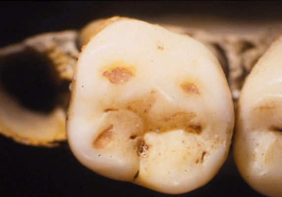
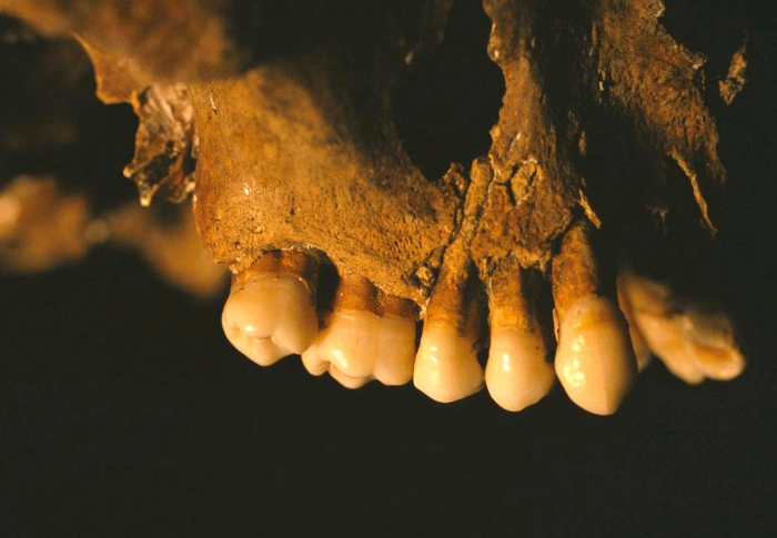
Figure 51: Photograph showing staining on the 2nd premolar and 1st molar on the right maxillary fragment belonging to Tom. |
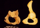
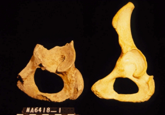
Figure 52: Photograph comparing part of the left hip bone belonging to Tom to a typical female hip bone (right side). |
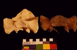
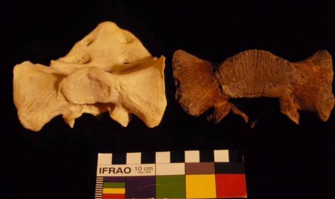
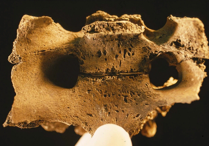
Figure 53:Photograph comparing the superior surface of the 1st sacral segment belonging to Tom to a typical female sacrum (left side). |
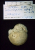
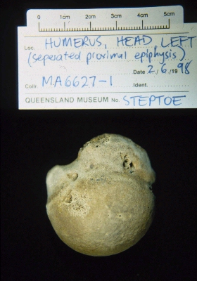
Figure 54:Photograph of a separated epiphysis of the head of the left humerus belonging to Tom. |
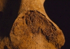
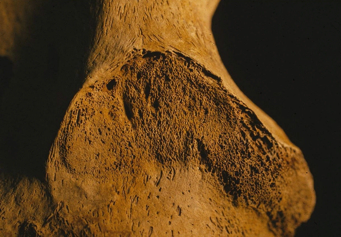
Figure 55: Photograph showing the auricular surface of the sacro-illiac joint on the right hip bone identified as belonging to Tom. |
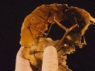
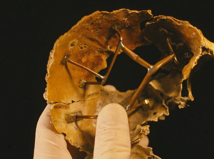
Figure 56: Photograph showing part of the reconstructed cranial fragments (frontal bone) belonging to Tom. |
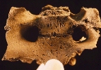
Figure 57: Pelvic surface of the 3rd and 4th sacral fragments belonging to Tom. Note that they are incompletely fused. |
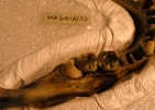
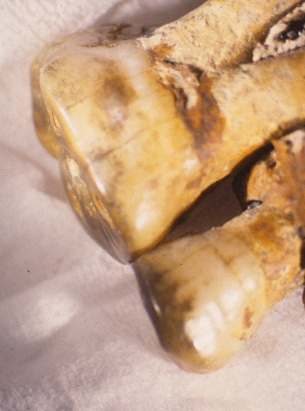
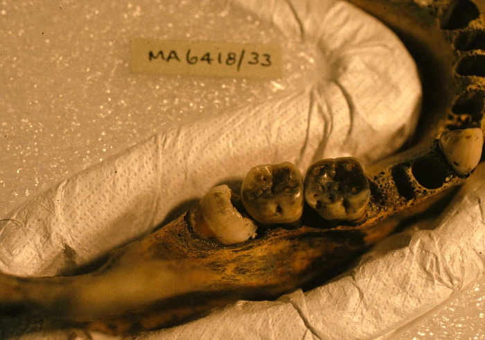
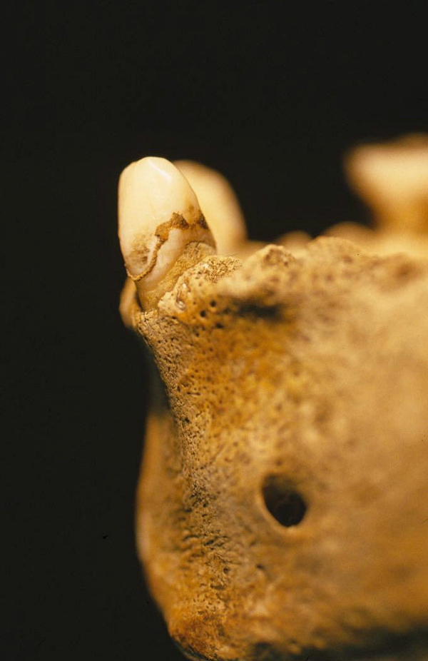
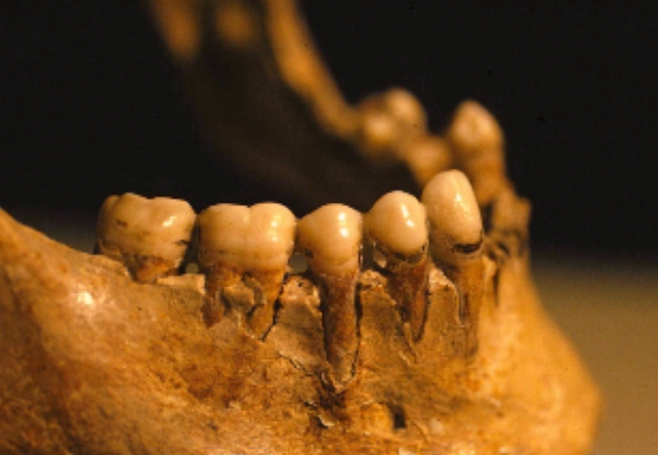
Figure 58: Right side dentition of Tom's mandible showing an impacted 3rd molar tooth. |
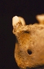
Figure 59:Photograph showing evidence of antemortem dental plaque on the dentition of Tom's mandible. |

Figure 60: Photograph showing evidence of antemortem dental plaque on the dentition of Tom's mandible. |
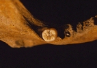
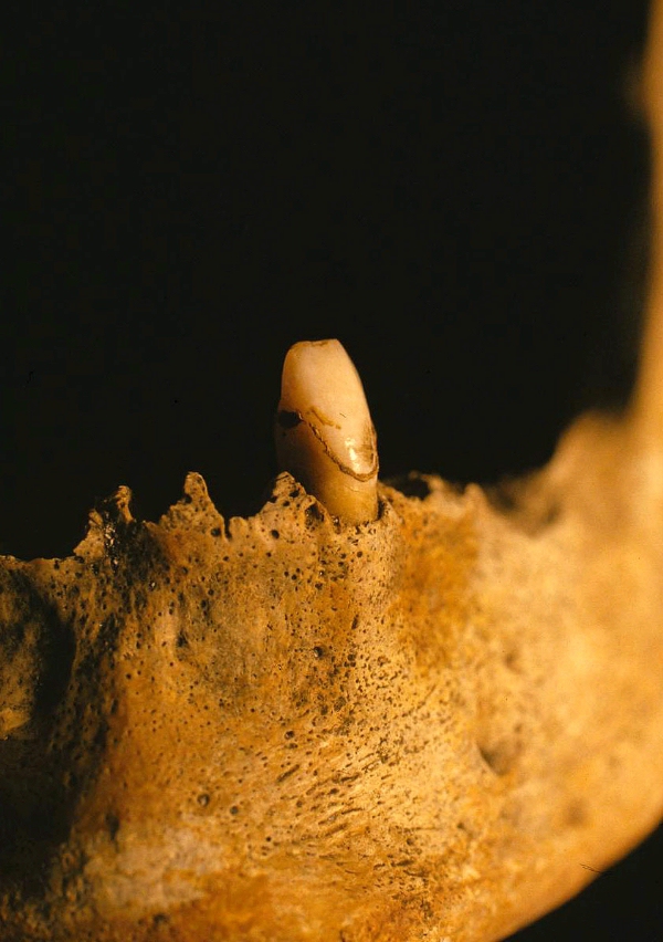
Figure 61:Photograph of the 38 tooth belonging to Tom. Note the absence of any obvious dental (dietary or occlusal) wearing. |
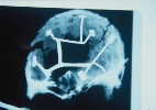
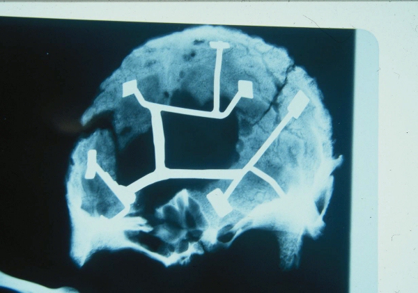
Figure 62:X-Ray showing some of the reconstructed portions of Tom's frontal bone |
| Other images of the remains of Dick |
|---|
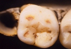
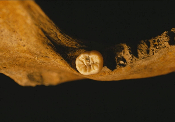
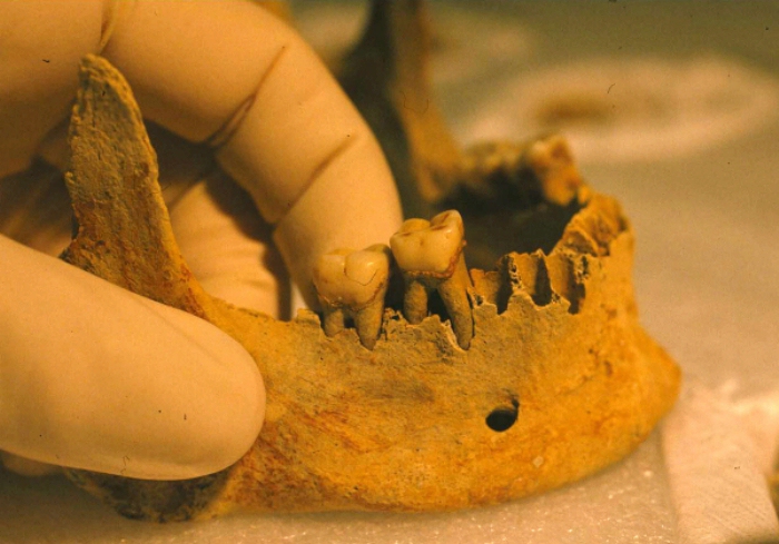
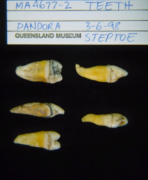
Figure 63: Molar tooth belonging from the mandible belonging to Dick. |
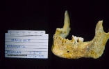
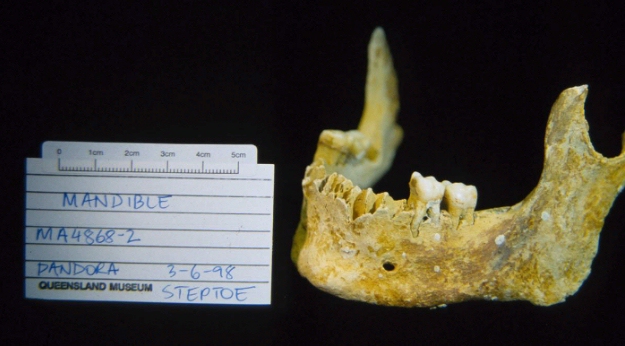
Figure 64: Dick's mandible. |
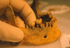
Figure 65: Dick's mandible. |
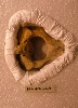
Figure 66: 2nd cervical vertebrae belonging to Dick. |
| Other images of the remains of Harry |
|---|
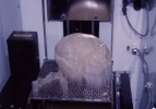
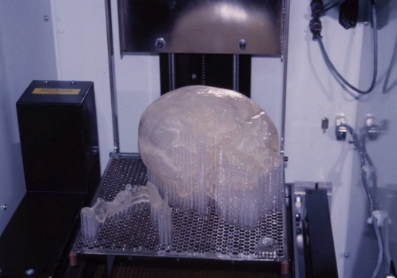
Figure 67: Harry's skull and mandible immediately after being duplicated by
stereo lithography. This skull was reconstructed by a series of over 750 horizontal slices of the original skull
scanned by a CT. |

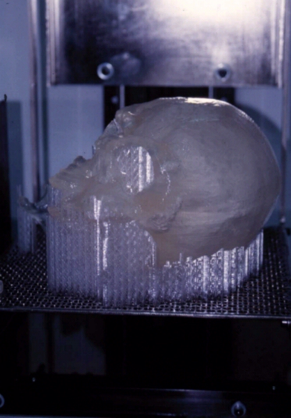
Figure 68: Harry's reconstructed skull. |
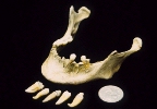
Figure 69:Harry's mandible. |
Figure 70:Photograph showing teeth belonging to Harry's skull. |
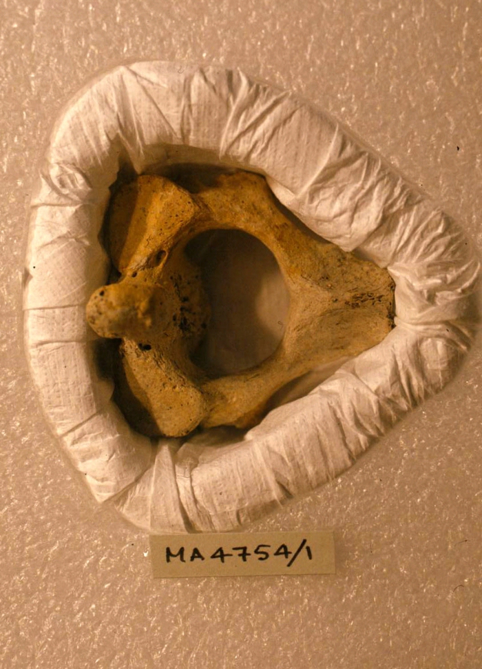
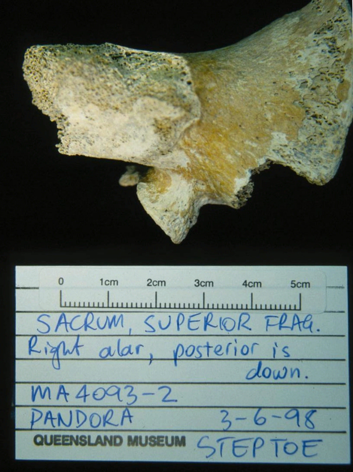
Figure 71:Superior view of a fragment of the first sacral segment. |
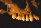
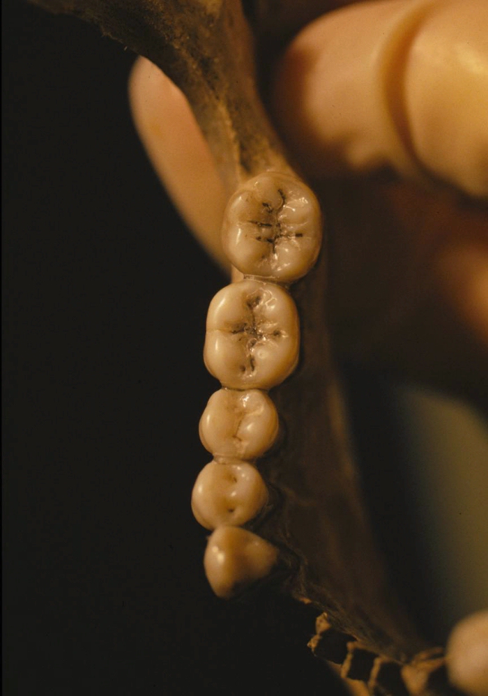
Figure 72:Photograph of the right maxillary dentition of Harry. Note the
exceptional condition of the teeth. |
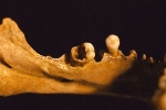
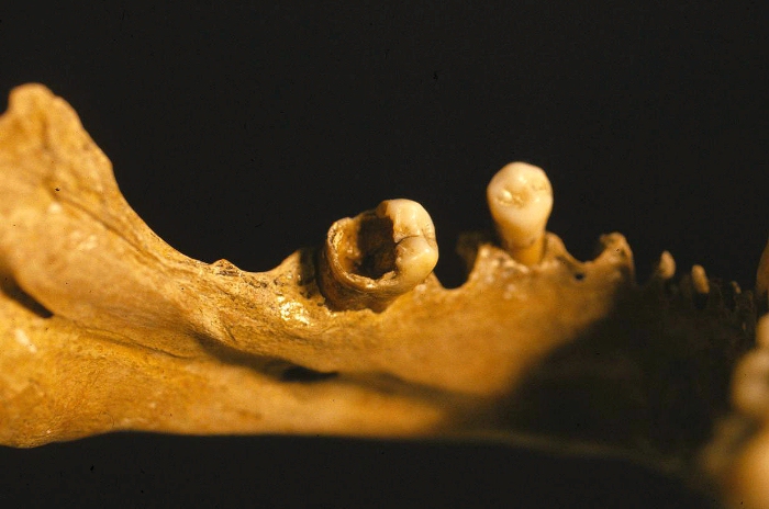
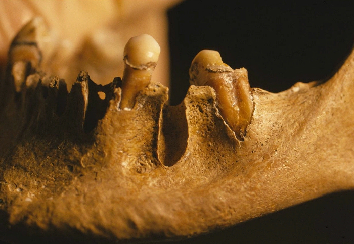
Figure 73: Photograph showing advanced dental infection of the 37 tooth of Harry's
mandible. |
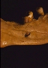
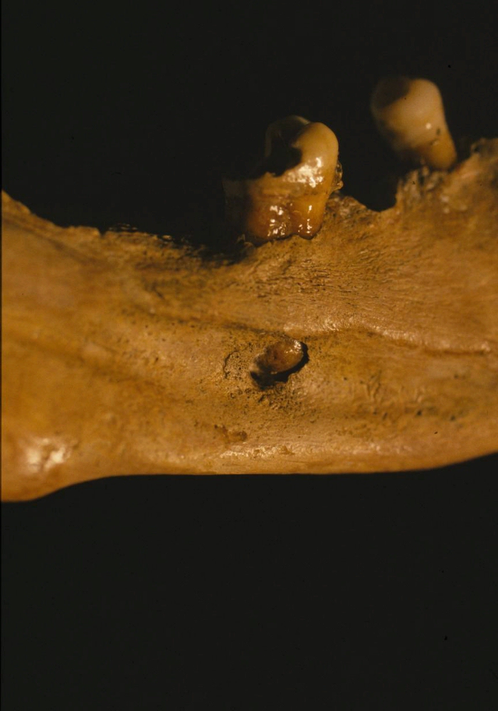
Figure 74: Medial view of the infection of the 37 tooth belonging to Harry's
mandible. Note the development of a bony abscess. |

Figure 75: Superior view of the right mandibular dentition belonging to
Harry. |
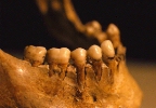
Figure 76:Right side mandibular dentition belonging to Harry. |
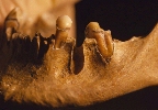
Figure 77: Photograph of the left side of Harry's mandible showing evidence of
periodontitis and antemortem plaque. |
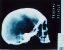
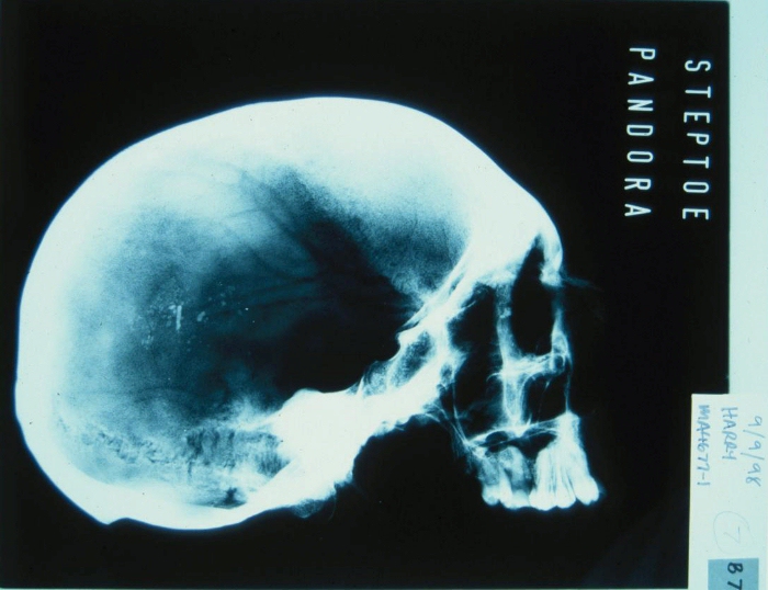
Figure 78: Lateral X-Ray of Harry's skull. |