Cite this as: Elliott, J. 2022 Radiographic Technique for Archaeological Human Dry Bones: a scoping review, Internet Archaeology 59. https://doi.org/10.11141/ia.59.1
Radiography has been used in the analysis of archaeological human skeletal remains to characterise or assist diagnosis of unknown pathologies, estimate age at death, demonstrate traumatic injuries and provide indications of biological stress (Mays 2007; Leo et al. 2013; Licata et al. 2019). In short, radiography facilitates the reconstruction of the biological profile of the deceased, although cultural modifications have also been explored (Ramírez-Salomón et al. 2018). In comparison with advanced imaging modalities such as computed tomography (CT), radiography is relatively inexpensive, more accessible and with lower logistical and training burdens (Garvin and Stock 2016; Vallis 2017). The advantages of CT cannot be dismissed though, with better visualisation of overlapping structures and generation of volumetric data allowing image reconstruction and interrogation (Beckett 2014). However, the lack of access to CT in commercial practice or academia is commonplace, with current British guidelines for recording human remains advocating radiography as a viable alternative (Mitchell and Brickley 2017). This study investigates the availability of literature guiding human dry bone radiography, primarily concerning the technical recommendations and workflow processes. In parallel with clinical use of radiography, this is termed radiographic technique. For the purposes of this study, archaeological human dry bones can be defined as complete or fragmented osteological remains lacking soft tissue typically recovered through excavation.
The application of radiography upon archaeological human dry bones has seen extensive interest, as demonstrated by several review articles and textbooks within academic literature (Conlogue et al. 2008; Chhem and Brothwell 2008; Beckett and Conlogue 2010; Beckett 2014; Licata et al. 2019; Beckett et al. 2020a; Conlogue and Beckett 2020). Furthermore, an abundance of research demonstrates its application in osteoarchaeological investigation such as tuberculosis (Évingera et al. 2011; Gooderham et al. 2020), osteogenesis imperfecta (Cope and Dupras 2011), and trauma (Bethard et al. 2021; Flensborg and Martínez 2021) among other conditions. The widespread use of radiography in osteoarchaeology is evident, and yet there is a lack of empirically based guidance for the act of imaging. Clinical (patient) radiography benefits from a plethora of instructional textbooks for patient management, image optimisation and radiographic technique (Whitley et al. 2015; Long et al. 2020). Nevertheless, the imaging of archaeological human dry bones presents unique challenges requiring a bespoke approach. For example, excavated remains may be incomplete and impregnated with soil or other debris (Elliott in press). Furthermore, disarticulated bones require an additional osteology skillset for accurate identification of laterality and orientation that may be unfamiliar to clinical radiographers (Elliott 2021). In order to overcome these challenges and develop a standardised approach for future research, an evidence-based solution based upon high-quality research is required. It is hoped that a unified approach formulated upon a robust methodology will allow comparable datasets to improve research potential.
This review sought to map existing literature related to human dry bone radiography within archaeology. Specifically, the aim of the study was to quantify and characterise current knowledge and recommendations related to radiographic technique or imaging workflow. Literature purely related to the interpretation of radiographic images fell outside the scope of this study. Radiography within this study relates to planar imaging involving the production of two-dimensional radiographic images (also known as 'plain film radiography'). This encapsulates digital systems, computed radiography or chemical film processing but excludes CT reconstructions or fluoroscopy.
A scoping review was adopted for this study owing to the heterogeneous nature of available literature, not restricted to time period of study (ancient-historic remains), research question or radiographic equipment deployed. The Arksey and O'Malley (2005) framework for scoping review methodology was used to inform the design of this study. Scoping reviews do not involve quality assessment of the literature as with systematic reviews, rather their goal is to map existing literature and answer broader research questions (Peters et al. 2015). Furthermore, the purpose of a scoping review is not to synthesise current knowledge to answer a specific question but to present themes and incidences of occurrence, often in graphical form. A comprehensive protocol with predefined objectives and methodology is required and outlined below.
Literature searches of JSTOR, PubMed and Science Direct were conducted using the search terms archaeology and radiography within the title or abstract, and paleoradiography, paleoimaging, or paleoradiology in any field. Publications were limited to academic textbooks or peer-reviewed journal articles written in the English language and published between 2001-2021. No geographical limitations were imposed; research was accepted from any country of origin. Relevant reference lists were also hand searched for additional literature. Searches were performed for each database on 1 November 2021.
| Inclusion criteria | Peer-reviewed literature or academic textbooks published between 2001-2021 in the English language |
|---|---|
| Radiography of human dry bones from any time period or geographical location of archaeological investigation | |
| Guidance or methodological assessment of radiographic technique relating to: Anatomy selected for imaging Radiographic views deployed Exposure factor selection Imaging workflow or concurrent activities | |
| Any medium of acquisition: computed, digital or wet-film processing | |
| Exclusion criteria | Non-human remains |
| Solely mummified remains | |
| Purely describing interpretation of radiographic imaging | |
| Below threshold for basic radiographic parameters: Exposure factors and radiographic views undertaken |
Study selection followed a three-stage process beginning with an assessment of title and abstract against eligibility criteria (Table 1). A broad inclusion of any study investigating or providing specific guidance for radiography of human dry bones were considered. Those involving non-human or mummified remains were excluded unless presenting information or results alongside human or disarticulated remains. The second stage involved reading the full text and applying a scoring system (Table 2) for progression onto stage three. Owing to the diversity in literature and iterative nature of scoping reviews some flexibility in methodology is acceptable (Peters et al. 2015). A scoring system was created in order not to risk excluding those articles attaining near-fulfilment of inclusion criteria despite clear application to human dry bones. Using this system, studies were either eliminated from the review, ascribed partial relevance or total relevance to the review aims (scoring one-three respectively). The scrutiny of study materials, methodology and concluding remarks acted as a failsafe check for inclusion prior to data charting. A threshold level for inclusion involved basic descriptions of technical radiographic details (exposure values, positioning) and/or specific commentary on bones selected for imaging. Any literature with a score of three was automatically progressed onto data charting.
| Score | Description and action taken |
|---|---|
| 1 | Not relevant Non-compliance with inclusion criteria. Eliminated from review |
| 2 | Partial relevance Article states use of radiography with human dry bones but does not specifically investigate or provide guidance for radiographic technique. Study materials, methodology and concluding remarks scrutinised for value to review |
| 3 | Total relevance Specifically investigates or provides guidance for radiographic technique of human dry bones. Included within review, progressed onto data charting for results. |
Data charting refers to the extraction of pertinent information from included articles to answer the review aim. The following information was extracted: type of literature, relevance to scoping review, application of radiography and recommendations specific to archaeological human dry bone. At its simplest, literature was divided between academic textbooks/peer-reviewed journal articles, with a further division between those articles that identified as primary data collection studies and guidance or protocol literature. Relevance to the scoping review included guidance or evaluation of technical specification for radiography (exposure values, specimen positioning or equipment setup), imaging workflow (bone selection, role division or concurrent activities) and quantitative analysis of bone (photodensitometry or radiogrammetry). The application of radiography within studies related to how imaging was used to answer specific research question(s) or objective(s). This was not always possible as academic textbooks covered a wide range of applications, and so were assigned as 'broad spectrum usage'. Lastly, included literature was scoured for specific recommendations for technical radiographic details or imaging workflow processes. For instance, the radiographic views undertaken (positioning of the specimen during imaging) or bone selection for pathologies. A predefined set of criteria for recommendations were not followed; instead an evolving list was generated in reaction to emerging publication themes.
Data were presented as a narrative thematic synthesis to quantify and characterise current research in human dry bone radiography. Critical appraisal of literature was not performed, as per the purpose of a scoping review.

A PRISMA 2020 (Page et al. 2021) flow diagram has been used to present the results of the literature search (Figure 1). Of the 244 unique journal articles or academic books identified using the search terms, 31 were obtained for full reading of text for assessment eligibility, with 21 publications subsequently satisfying the inclusion criteria (Appendix). A literature map is presented in Figure 2, outlining the current research and academic textbooks for archaeological human dry bone radiography based upon the search results of this study.
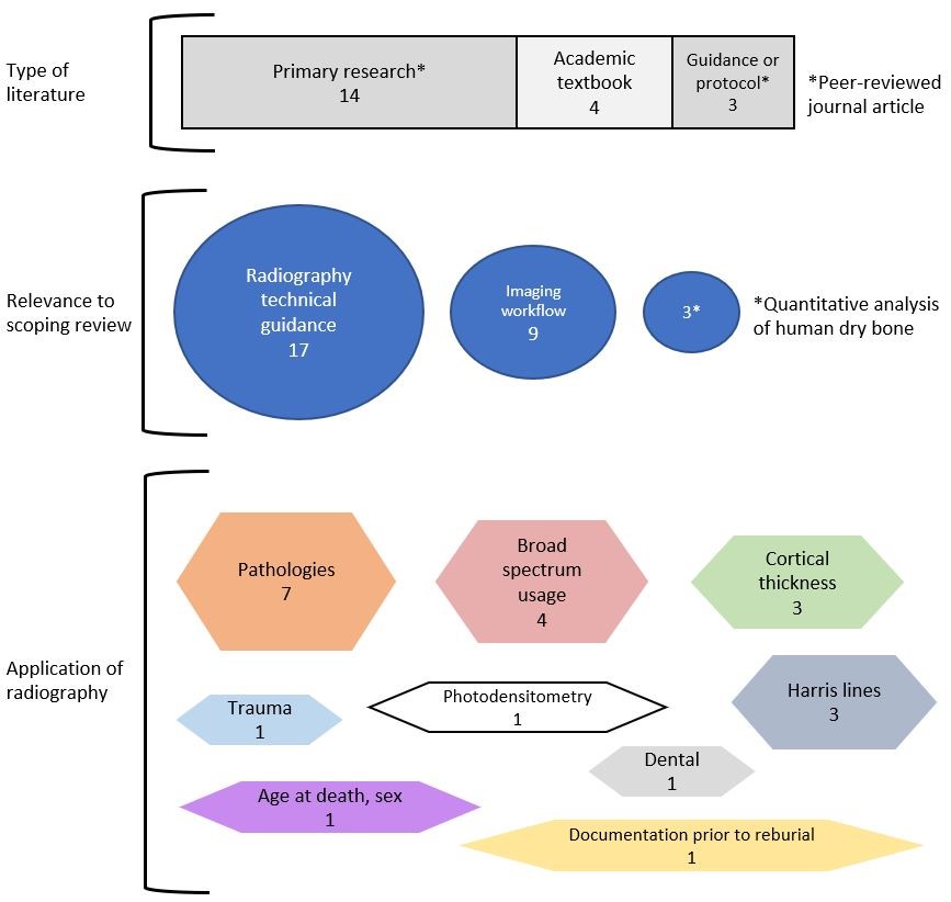
Peer-reviewed journal articles (n = 17) were found to be more prevalent than academic textbooks (n = 4). Of those articles, primary research (n = 14) were more common than guidance or protocol publications (n = 3). While categorising type of literature was simple, relevance to the scoping review was less clear, with some literature contributing to multiple facets. The majority of publications (n = 17) were relevant to technical aspects of radiography, with nine providing recommendations for imaging workflow. A small portion of the literature provided direct procedural guidance for quantitative analysis of archaeological human dry bones (n = 3), including details on bone selection and/or radiographic views necessary.
A myriad of research objectives were identified during the scoping review, as shown in Figure 2. Nevertheless, the primary focus was upon the investigation of pathologies (n = 7), cortical thickness as an indicator of bone loss (n = 3) and Harris Line investigation (n = 3). Academic textbooks lacked a singular research objective per se, instead providing the foundational knowledge required for the application of radiography. For instance, Beckett and Conlogue (2010) and Conlogue and Beckett (2020) include radiographic theory alongside practical examples of application. These case studies present unique challenges, solutions and recommendations (particularly for field applications). Not all case studies involved dry human bone though, with a preponderance of mummy examples; however the core concepts were relevant.
Of the 21 studies included within this review, only three self-identified in their title as guidance or protocol for radiographic imaging. The remainder either offered a general overview of the topic, as with academic textbooks, or specific recommendations due to primary data collection findings. An excellent example of the latter includes Primeau et al. (2016), with their investigation into Harris Lines leading to evidence-based recommendations for radiographic views. In contrast, other primary research studies provided detailed methodological accounts of radiography but with an absence of specific recommendations in the conclusion or elsewhere. Such studies hold value though, as with Biehler-Gomez et al. (2019), whose extensive photographic and radiographic figures may serve as indicative examples of technique for future studies. Table 3 provides a break-down of recommendations for radiography of archaeological human dry bone. Recommendations for X-ray exposure factors, radiographic views and bone selection for imaging were most prevalent whereas specialist procedures such as radiogrammetry or photodensitometry were lacking. Overall, there were more publications addressing radiographic technical details than imaging workflow, but these addressed the wider interaction with affiliated disciplines such as anthropology, osteoarchaeology (both for macroscopic inspection) and radiology (for image interpretation).
| Radiographic technical details | Literature |
|---|---|
| Exposure selection (e.g. tube current or voltage) | 2, 4, 6, 8, 16, 17, 19 |
| Postcranial radiographic views | 1, 2, 9, 15, 18, 19, 21 |
| Cranial views | 2, 5, 7, 14, 16, 18, 20 |
| Dental (mandible/maxilla) views | 5, 12, 16 |
| Radiogrammetry process | 2, 3, 21 |
| Photodensitometry process | 8 |
| Advocates clinical radiographic views and positioning | 4, 9 |
| Imaging workflow | Literature |
| Bone selection for systematic documentation | 1 |
| Bone selection for survey of specific pathologies | 10, 11, 13, 15, 18, 20 |
| Bone selection for photodensitometry | 8 |
| Equipment/facilities set up | 4, 6, 17 |
| Proformas for imaging process | 6, 17 |
| Team role workflow diagrams | 18, 19 |
| Advocates photography as complementary imaging | 1, 6, 15, 16, 18 |
Although sparse, the results of this scoping review demonstrate the existence of literature specifically catering for radiographic technique with archaeological human dry bone. Furthermore, a plethora of applications have been identified, albeit in a relatively small number of publications. It should be reiterated that the results do not simply represent the use of radiography within archaeological literature, but the evaluation or recommendations for its application. A full account of all literature involving radiographic analysis would be of little value, except perhaps validating its use in archaeological practice. A variety of publications directly addressed radiography as an imaging modality in archaeology but did not offer practical advice and were therefore excluded from the review (Chhem 2006; Beckett 2014; Licata et al. 2019; Wanek et al. 2021). In these examples the authors offer well-informed evaluations of radiography as an imaging modality or are dedicated to the interpretation of trauma or pathologies on radiographs but do not provide recommendations, as shown in Table 3. The inclusion of photogrammetry or radiogrammetry articles within the results may come under scrutiny, as they align closer to image interpretation than acquisition. However, the procedural nature and practical advice concerning bone selection and/or radiographic views warranted their acceptance. For instance, Manifold (2014) suggests excluding bones with disease, trauma or soil infiltration during photodensitometry. Other literature was tantalisingly close to inclusion but was relegated because of involvement with mummies (Kristóf et al. 2015; Beckett et al. 2020b), or animal remains (Symmons 2004). The exception was Seiler et al. (2018), who gave an account of both mummified and skeletonised human remains.
A key factor for exclusion was the omission of basic radiographic parameters within methodologies. This was more noticeable with primary research studies where radiography was pivotal to their research goal and yet details were scant. To illustrate the point, an assortment of archaeological investigations for disease, biological stress, bone loss or trauma yielded excellent examples of radiography in practice but lacked adherence to the review aims (Ameen et al. 2005; Dabernat and Crubézy 2009; Beauchesne and Agarwal 2017; Cieślik et al. 2017). Conversely, publications were found that directly addressed radiographic methodology, which may assist standardisation of future practice. Bruwelheide et al. (2001) present a detailed protocol for radiographic and photographic documentation of remains prior to reburial or repatriation. Specifically, the authors provide both visual and written explanations of specimen selection and positioning for imaging, accompanied by examples and recommendations for broad-based documentation. The texts of Beckett and Conlogue (2010) and Beckett et al. 2020a) offer a host of practical considerations, including proformas for recording specimen imaging and example risk assessments. Lastly, Elliott (2021) and Meyer et al. (2020) provide workflow diagrams demonstrating the transit of specimens between team member specialisms (e.g. photography, radiography, osteology) and the documentation process.
Although the results demonstrate a wide range of applications of radiography, the paucity of literature indicates that greater investigation is required to inform practice. The majority of available research concerns the identification of pathologies, leading to the assumption that this represents the foremost area of enquiry within osteoarchaeology. Bone loss, whether volume or density, has seen concerted research efforts (see Mays 2016; Agarwal 2018) but requires greater guidance at a practical level to encourage or facilitate further investigations. The recent article by Gilmour et al. (2021) concerning metacarpal radiogrammetry may serve as an exemplary format. The authors include a detailed radiographic methodology, with accompanying procedural guidelines for quantitative analysis of the resultant imagery. An evidence-based approach using a skeletal collection of known provenance allows for greater reliability and more robust recommendations as a result. Similar studies for other regions of anatomy, especially regarding photodensitometry, would be beneficial.
Interestingly, several studies refuted the value of radiography to identify bone lesions, stating that macroscopic osteological analysis (by eye) is superior (van Schaik et al. 2017; 2019). However, Fatula (2021) clarifies the issue, stipulating that occult lesions that are invisible to the eye may only be identified through radiographic means (if a non-destructive approach is desired). A pragmatic approach would therefore include visual inspection followed by radiographic imaging for confirmation of diagnosis or a skeletal survey to account for other pathological manifestations elsewhere (i.e. metastasis, congenital or metabolic malformations). Bruwelheide et al. (2001) and Biehler-Gomez et al. (2019) provide the strongest arguments for radiography and photography to be used in tandem as complementary methods of recording the deceased. Literature regarding the photography of human dry bone undoubtedly exists, but the integration with radiography requires further investigation.
This scoping review was limited to archaeological literature, thereby excluding potentially aligned disciplines that may offer valuable insights. For instance, radiography has been utilised during victim identification in forensic investigations of skeletonised remains to estimate age through dental eruption analysis (Ashifa et al. 2020). Other studies, such as Silva et al. (2013), present the use of radiography with dry bones to match post-mortem and ante-mortem dental and sinus appearances. Although incongruent with the end-purpose of the imaging (i.e. to answer questions of the law), archaeologists may yet learn valuable lessons concerning methodology, logistics and interdisciplinary collaboration. Another limitation concerns the exclusion of non-English language publications, which may have introduced language bias, thereby potentially eliminating valuable literature. Lastly, the rapid digital access to journal articles was in contrast to a slow process of inter-library loans for textbooks, potentially limiting their inclusion within this study.
A total of 21 publications have been found that provide guidance, protocols or recommendation for radiography of archaeological human dry bones. The majority of these are peer-reviewed journal articles, dominated by primary research studies. Radiography has been applied to a wide variety of research objectives including identification of pathologies, Harris Lines and visualisation of trauma. Literature based upon primary data collection tended to provide recommendations for specific tasks, whereas academic textbooks were found to have a wealth of case studies and details regarding equipment or documentation process. Excluded literature typically lacked the methodological detail or subsequent recommendations to be of value to this review, despite direct relevance to human dry bones. Future research may benefit from a holistic approach to human dry bone remains, incorporating both photography and forensic research to support a standardised approach.
| Number | Author(s) | Year | Title |
|---|---|---|---|
| 1 | Bruwelheide et al. | 2001 | Standardized protocol for radiographic and photographic documentation of human skeletons |
| 2 | Nystrom et al. | 2004 | Field paleoradiography of skeletal material from the Early Classic period of Copan, Honduras |
| 3 | Ives and Brickley | 2004 | A procedural guide to metacarpal radiogrammetry in archaeology |
| 4 | Conlogue et al. | 2004 | The application of radiography to field studies in physical anthropology |
| 5 | Chhem and Brothwell | 2008 | Paleoradiology: Imaging mummies and fossils |
| 6 | Beckett and Conlogue | 2010 | Paleoimaging: field applications for cultural remains and artifacts |
| 7 | Papagrigorakis et al. | 2012 | Paleopathological findings in radiographs of ancient and modern Greek skulls |
| 8 | Manifold | 2014 | Photodensitometry: a useful method for studying bone mineral density in the skeletal remains of children |
| 9 | Scott and Hoppa | 2015 | A re-evaluation of the impact of radiographic orientation on the identification and interpretation of Harris Lines |
| 10 | Primeau et al. | 2016 | CT imaging vs. traditional radiographic imaging for evaluating Harris Lines in tibiae |
| 11 | van Schaik et al. | 2017 | The radiologist in the crypt: Burden of disease in the past and its modern relevance |
| 12 | Seiler et al. | 2018 | Application of portable digital radiography for dental investigations of ancient Egyptian mummies during archaeological excavations: Evaluation and discussion of the advantages and limitations of different approaches and projections |
| 13 | van der Merwe et al. | 2018 | Four possible cases of osteomalacia: the value of a multidisciplinary diagnostic approach |
| 14 | Purchase et al. | 2019 | An innovative method to visualise mastoiditis using a hand-held X-ray system |
| 15 | Biehler-Gomez et al. | 2019 | The synergy between radiographic and macroscopic observation of skeletal lesions on dry bone |
| 16 | Beckett et al. | 2020a | Case studies for advances in paleoimaging and other non-clinical application |
| 17 | Conlogue and Beckett | 2020 | Advances in paleoimaging: applications for paleoanthropology, bioarchaeology, forensics, and cultural artifacts |
| 18 | Meyer et al. | 2020 | Multidisciplinary studies of heavily fragmented and commingled ancient Egyptian human remains found in KV 40 (Valley of the Kings, Luxor, Egypt): a pragmatic workflow and first results |
| 19 | Elliott | in press | Radiography of human dry bones: a reflective account with recommendations for practice |
| 20 | Fatula | 2021 | Detection of cancerous lesions in skeletal remains using visual methods and radiographs |
| 21 | Gilmour et al. | 2021 | Quantifying cortical bone in fragmentary archeological second metacarpals |
This project has been funded by the College of Radiographers Industry Partnership Scheme Research Grant (Grant number 186, 2020).
I'd like to thank the staff of Maidstone and Tunbridge Wells NHS Trust and Canterbury Christ Church University for supporting me during the creation of this research.
Internet Archaeology is an open access journal based in the Department of Archaeology, University of York. Except where otherwise noted, content from this work may be used under the terms of the Creative Commons Attribution 3.0 (CC BY) Unported licence, which permits unrestricted use, distribution, and reproduction in any medium, provided that attribution to the author(s), the title of the work, the Internet Archaeology journal and the relevant URL/DOI are given.
Terms and Conditions | Legal Statements | Privacy Policy | Cookies Policy | Citing Internet Archaeology
Internet Archaeology content is preserved for the long term with the Archaeology Data Service (ROR). Help sustain and support open access publication by donating to our Open Access Archaeology Fund.
 Home
Home