Through much of history childhood was a risky time, and one that was probably punctuated by periods of nutritional shortage and/or illness. Where the child survives the stress, 'markers' may remain on the teeth and/or bones, and the age at which the upset occurred can be estimated. Horizontal grooves or pits on the teeth — enamel hypoplasia — represent 'upsets' which occurred whilst the tooth was developing, whilst lines of increased density in the long bones reflect the recovery phase of illness. The latter is examined by section or radiographically and as such is beyond the scope of the present study. However, Cross (1988) conducted a detailed study of lines of increased density in the 1980-1 material and her findings, which it seems likely would equally apply to the 1994 sample, are summarised below.
Nutritional deficiency
A number of vitamin deficiencies leave evidence on the bones. These include deficiencies of Vitamins D (leading to rickets or osteomalacia) and C (leading to scurvy). There were no examples of either of these conditions in the present sample. Rickets did not become common until after the industrial revolution when much of the population received little exposure to sunlight, preventing the body from synthesising its own Vitamin D. Scurvy is common when there is little access to fresh fruit and vegetables, and whilst the late medieval population periodically may have had restricted access to food, it seems likely that fresh green vegetables were generally plentiful, preventing such deficiencies. Iron deficiency anaemia can be detected from the bones.
Cribra orbitalia
Pitting in the roof of the eye socket known as cribra orbitalia is usually thought to indicate childhood iron deficiency anaemia (Stuart-Macadam 1985). The deficiency may be of dietary origin but is perhaps more likely to reflect a high disease/pathogen load. Pathogens may cause iron deficiency anaemia directly, as in the destruction of red blood cells in malaria and in blood loss from the intestines in hookworm infestation, or indirectly as the body may reduce the amount of available iron in the blood serum as a generalised response to stress. In this way iron deficiency anaemia may confer some protection against disease as many micro-organisms need iron from their 'host' in order to replicate (Stuart-Macadam 1992). The picture is further complicated by scurvy and infections of the eye, both of which may produce similar lesions. The three may be impossible to distinguish in the archaeological context.
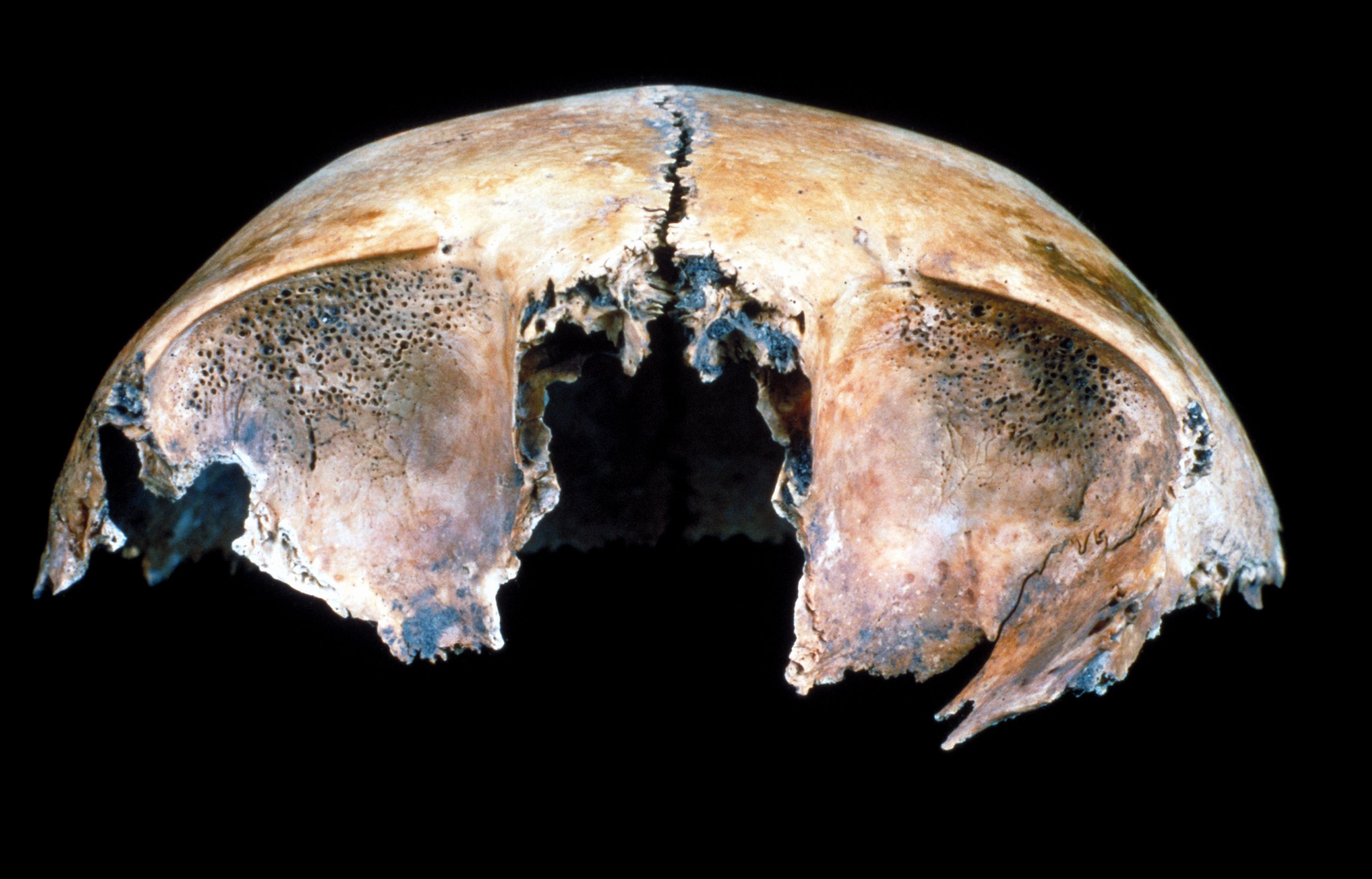
Ten percent of adults (5/48) and 27% of immatures (6/22) displayed cribra orbitalia [Photo 0131], most being of a minor degree (Table 33). Variation in the frequency of cribra with age is seen in many other sites and may indicate higher mortality in children that developed pitting, healing and obliteration of lesions with age, or more probably a combination of the two. A comparison of stature between groups with and without cribra was attempted but unfortunately the sample size of adults with both cribra and an estimate of stature was too small to provide meaningful data.
Seven percent of adults (7/96) and 34% of immatures (16/47) displayed cribra orbitalia. The distribution by sex was not examined due to the small sample size. Similarly, comparison of stature between groups with and without cribra was not made because the sample size of adults with both cribra and an estimate of stature was too small.Enamel hypoplasia
Enamel hypoplasia is a condition which results in defects such as lines, pits or grooves on the enamel surface of the teeth. These defects occur whilst the teeth are developing and remains as a permanent record into adulthood. They are caused by nutritional deficiency or childhood illnesses such as measles. Preliminary analyses suggest that more than half of immatures (57%; 4/7) and four fifths of adults (82%; 18/22) displayed enamel hypoplasia, which was minor in most cases. Enamel hypoplasia is most commonly observed on the permanent dentition and the discrepancy between age groups probably reflects the small number of permanent teeth in the immature group.
Harris Lines
Harris lines are lines of increased density seen in the long bones that are most commonly examined on x-radiographs. They are thought to represent the recovery phase following a period of growth disturbance such as may have been caused by illnesses such as measles or whooping cough or by malnutrition and as such are considered to be non-specific markers of biological insult. The lines may persist into adulthood although it seems probable that some are lost as the bones are remodelled throughout life.
Ninety-five adult and immature tibiae (1980-1 sample) showed that 89% of immatures and 91% of adults displayed at least one Harris line, immature tibiae displaying on average 5.6 lines, whilst adult tibiae displayed 3.3. Whilst the high frequency of Harris lines indicates a population subject to frequent stresses, it also indicates that the stresses were not chronic as long term stress prevents the recovery phase and inhibits the development of Harris lines (Murchison et al. 1984, Steinbock 1976, in Cross 1988). Cross therefore concludes that 'the Aberdeen sample appears to have suffered — and recovered — from relatively frequent episodes of growth disturbance', possibly of a seasonal nature (Cross 1988, 157).
Osteoporosis
Osteoporosis is caused by an imbalance of bone resorption and bone production and causes bones of decreased density which are fragile and liable to fracture with minimal trauma. It may be associated with disease (e.g. Cushing's syndrome), medical treatment (e.g. long-term steroid therapy) old age (senile osteoporosis) or other, unknown factors (idiopathic osteoporosis). Most archaeological examples can be assumed to represent the senile form, the changes occurring rapidly in women after the menopause and much more gradually in men. Osteoporosis is seen most often in women after the 5th decade and in men after the age of seventy (Roberts and Wakely 1992). Osteoporotic bones tend to fracture in areas with a large proportion of cancellous to cortical bone and consequently of most rapid turnover, such as the vertebral bodies and the femoral neck. Vertebrae may become dished and compressed, causing a decrease in stature and dorsal kyphosis ('dowager's hump', seen most often in elderly women). Femoral neck fractures tend to occur approximately 15 years later than vertebral fractures (Revell 1986 quoted in Miles 1989) and are difficult to heal. Thirty percent of beds in modern orthopaedic wards are occupied by those with fractures associated with osteoporosis (National Osteoporosis Society 1993).
Six cases of osteoporosis were seen in the present population (information from 1994 excavation only). Whilst there were no fractures of the femoral neck or vertebral bodies, the condition was indicated by light, fragile bones in six skeletons. In one case (SK 223) this may have been related to leprosy infection/psoriatic arthropathy. Four of the five remaining cases were female, indicating that these individuals lived beyond the menopause and were probably over fifty years old. Three of the females were classified as late middle aged, the other as middle aged. As osteoporosis is associated with low levels of osteoarthritis (A Stewart, pers. comm.) an under-ageing of the mature female population in particular may be indicated when degenerative change is used as one of the principle ageing criteria.
Sero-negative arthropathies
Sero-negative arthropathy including rheumatoid arthritis, ankylosing spondylitis, psoriatic arthritis and Reiter's syndrome may leave evidence on the bones. Diffuse idiopathic skeletal hyperostosis (DISH) is included here as it shares a number of features with the sero-negative arthropathies although its aetiology is unknown. The common features of this group of disorders is fusion of spinal elements, often with involvement of the sacro-iliac joints, development of enthesopathies around the pelvis and the heel and a tendency for males to be affected more often than females.
No cases of rheumatoid arthritis were identified. It has been suggested that this disorder is a modern disease as it affects 3% of females and 1% of males today (Anderson 1985) but is rarely identified in archaeological groups. However the disease is associated with osteoporosis, fragile bones and entirely lytic lesions around the joints. These features combine to make survival of affected bones and recognition of lesions unlikely in many archaeological situations.
There was no clear evidence of any other sero-negative arthropathy. The disarticulated left innominate and sacrum of an adult male (context 964) were fused at the sacroiliac joint, but the sacral-lumbar joints which would tend to be affected in sero-negative arthropathy were uninvolved. SK 223 may have suffered from psoriatic arthritis, a form of sero-negative arthropathy. Differential diagnosis includes leprosy and this individual is discussed above.
One possible example of DISH (diffuse idiopathic skeletal hyperostosis) was noted. SK 221 (old male) displayed typical fusion of the bodies of three thoracic vertebrae, enthesopathies of the ischial tuberosity, iliac crest, greater and lesser trochanters, gluteus maximus, bicipital tuberosity of the radius, brachialis insertion of the ulna and distal tibia/fibula articulation. Multiple cystic lesions of the hand and distal tibia were also noted and were probably related to osteoarthritis. DISH is thought to affect 4% of males and 3% of females over 40 years and 10% of men and 7% of women over 60 years (Ehrenfeld 1990). The gross appearance of this individual indicates a middle-aged individual although the radiographic appearance is of an elderly man, probably in his 70's (J. Weir pers. comm.). This again may indicate a general under ageing of the older age groups of skeletal populations.
Developmental/Congenital
Variation in vertebral number/form: There were four examples of extra vertebrae, six sacral vertebrae in SK 230 and six lumbar vertebrae in SK 245, SK 256 and SK 257. No transitional vertebrae were seen. The anomalies are unlikely to have had any clinical significance. Apparent congenital fusion of the vertebral bodies or arches was seen in two individuals (Cross 1988, 100).
Spina bifida: Spina bifida is characterised by incomplete fusion of the posterior vertebral elements, usually in the lower spine. It is largely limited to the sacrum in archaeological populations, as those affected in other areas probably did not survive. There were two examples of complete sacral spina bifida (3.6%, 2/55), both from the 1980-1 excavation (Table 34). Frequencies at other locations on the sacrum are shown in the table. The 5th sacral vertebra was frequently open and this was considered a normal variant. It is noteworthy that all examples of deficient fusion come from the graveyard to the east of the church with two of the affected individuals being side by side and both displaying metopism.
Internet Archaeology is an open access journal based in the Department of Archaeology, University of York. Except where otherwise noted, content from this work may be used under the terms of the Creative Commons Attribution 3.0 (CC BY) Unported licence, which permits unrestricted use, distribution, and reproduction in any medium, provided that attribution to the author(s), the title of the work, the Internet Archaeology journal and the relevant URL/DOI are given.
Terms and Conditions | Legal Statements | Privacy Policy | Cookies Policy | Citing Internet Archaeology
Internet Archaeology content is preserved for the long term with the Archaeology Data Service. Help sustain and support open access publication by donating to our Open Access Archaeology Fund.