A number of miscellaneous conditions were noted:
SK 214, a middle-aged male, displayed slight flattening and porosity of the lunate facets of both radii and a small, distorted, pitted lunate. The aetiology of this condition is unknown.
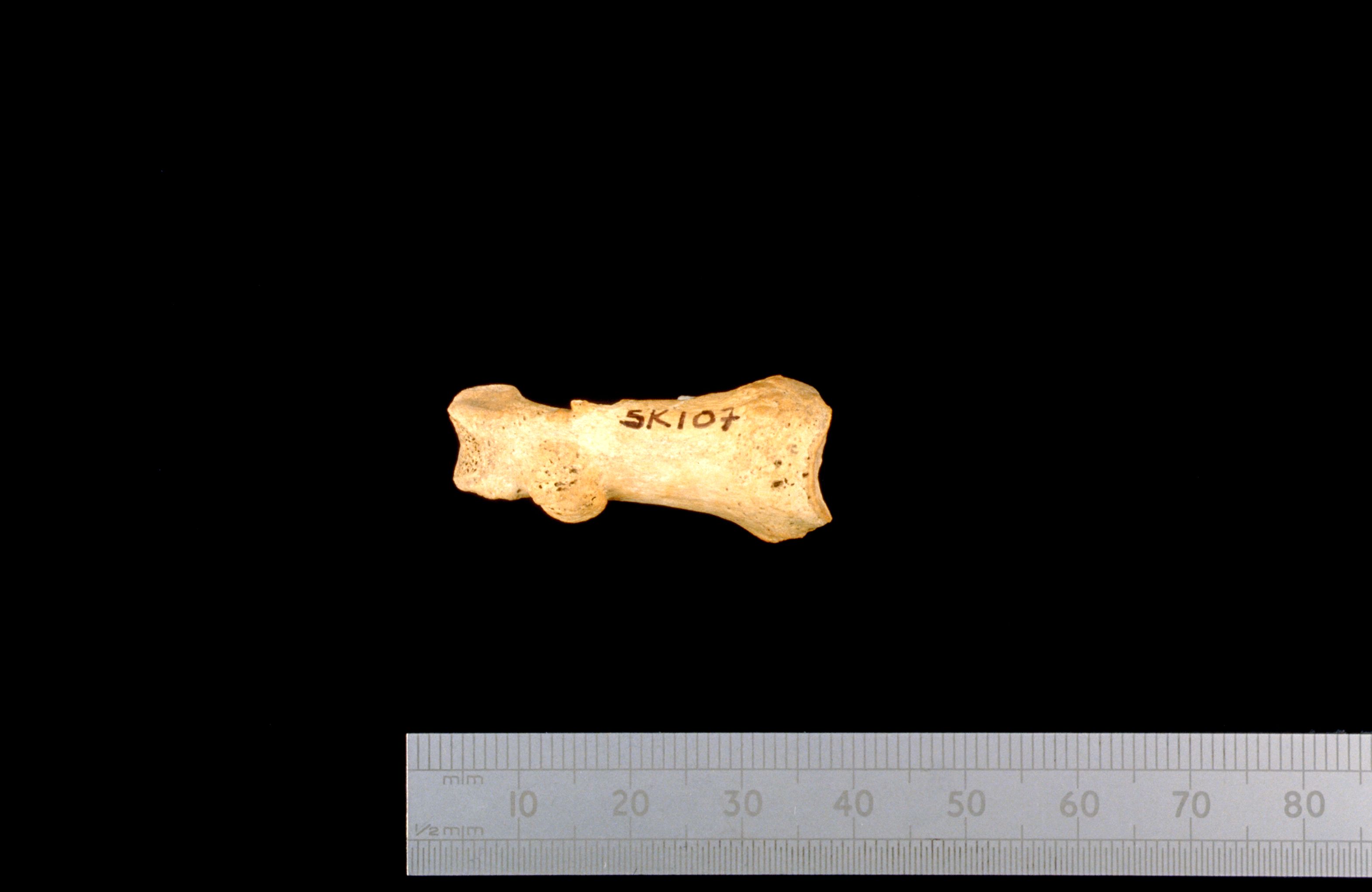
SK 307, a middle-aged possible male, displayed a benign-looking, rounded deposit of bone [Photo 0129], 5 mm in diameter, on the dorsal surface of one proximal hand phalanx. The quality of the bone appeared to be normal and there was no evidence of infection or other pathology.
A spherical object with a bone-like appearance, 9 mm in diameter, probably a bladder or kidney stone, was found amongst the disarticulated material (context 1210).
A number of immature rib fragments was found in contexts 708 and 916 although common features suggest that they come from the same individual. The bones were slightly misshapen — as though made of plasticine with depressions caused by finger prints — with new bone alongside circumscribed lytic lesions and a general porosity of the bones. Radiography revealed very thin cortices and an unusual trabecular pattern, but consultation with clinicians failed to identify the aetiology.
The ulna of SK 324, whilst severely eroded, displayed an unusual degree of curvature. Radiography revealed extensive new bone formation over the entire length of the shaft. The skeleton was very incomplete and no other pathology was noted.
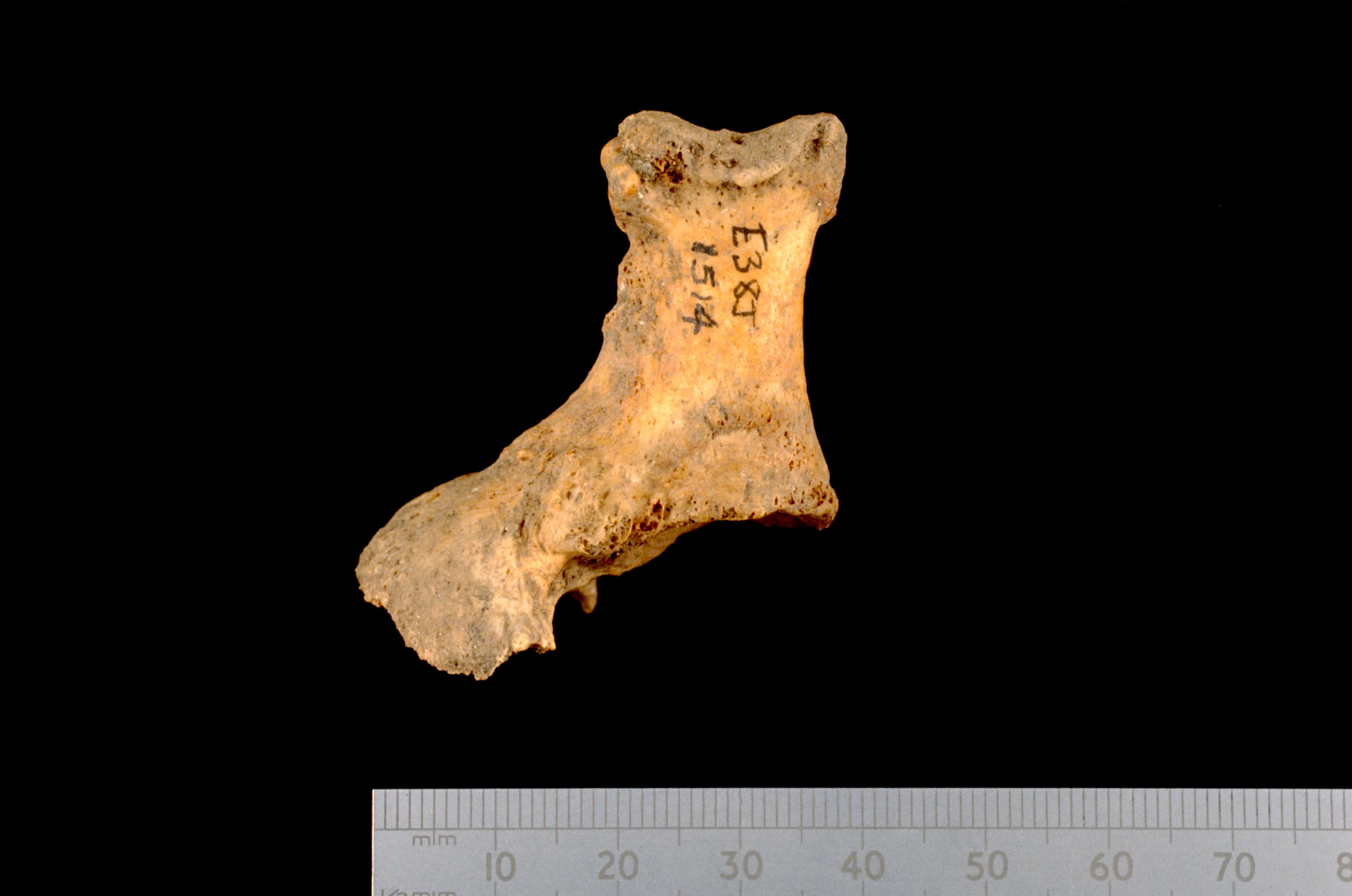
The disarticulated proximal phalanx of the great toe of an adult (context A1514) displayed a large lateral growth at the proximal extremity [Photo 0126]. The articular surface was unaffected. The growth probably represents ossification into the ligaments as a result of continued trauma. The growth would have produced a prominent, hard lump on the medial side of the foot which may have looked somewhat like a bunion.
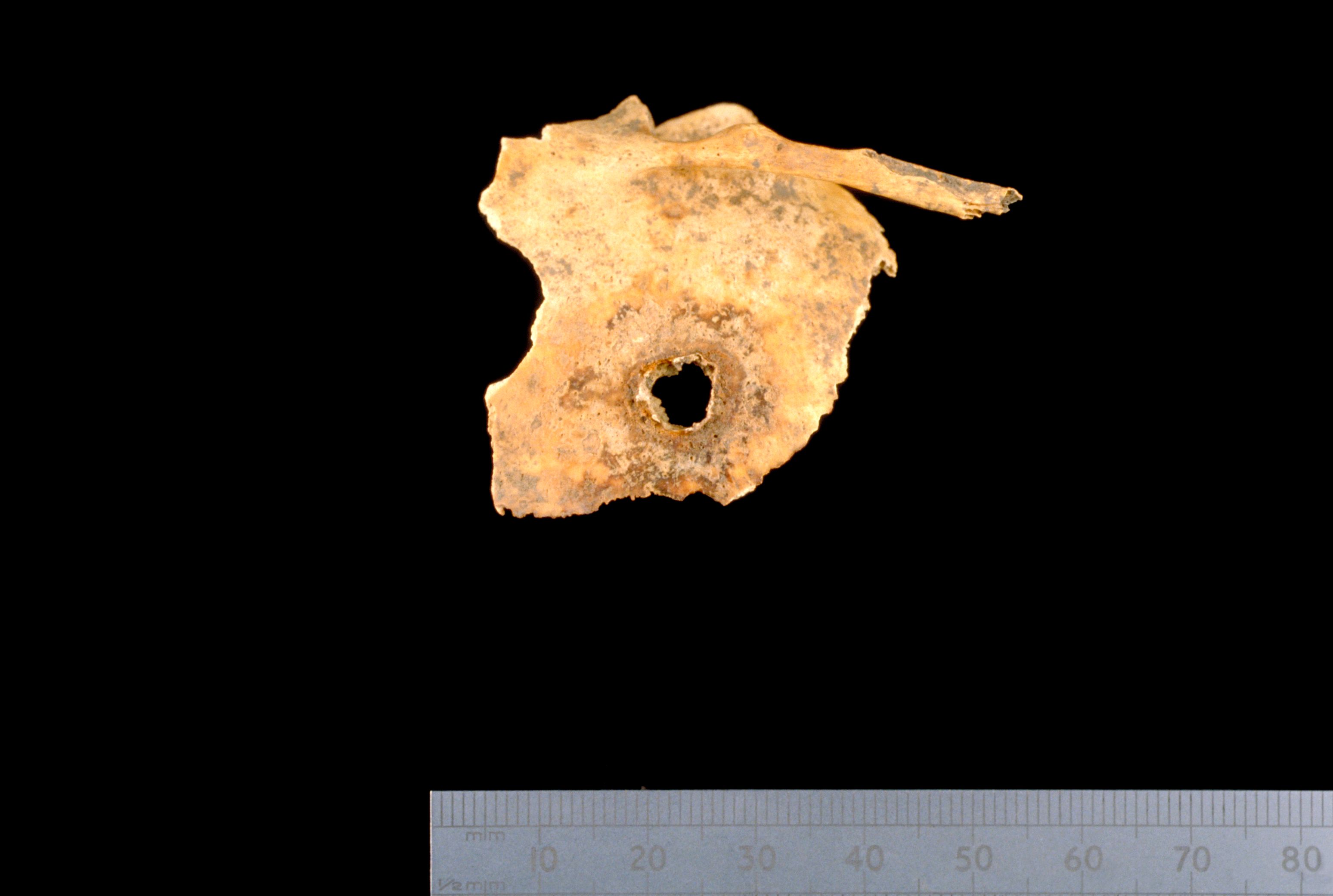
A 4 mm lesion [Photo 0128] perforated the left temporal of a child aged 5–6 years (SK 216). There was a slight build-up of bone with porosity present around the margins of the lesion and fine, unhealed periosteal new bone on the internal surface of the cranium. The aetiology of the lesion remains elusive despite consultation with clinicians, possible explanations including a perforating injury, or infection of the bone by a blood-borne organism.
The medial condyle of the left femur of a female displayed a large depressed area which superficially resembled a healed fracture, although the site of the defect and the lack of radiographic evidence indicate that this diagnosis is unlikely (SK 235). An alternative explanation is of an atypical osteochondritis dessicans (Weir/Chesney, pers. comm.).
A disarticulated humerus displayed a pronounced groove of the distal articular surface (context 1211). There was no radiographic evidence of fracture and the feature may reflect trauma during infancy or be of developmental origin (J. Weir, pers. comm.).
Internet Archaeology is an open access journal based in the Department of Archaeology, University of York. Except where otherwise noted, content from this work may be used under the terms of the Creative Commons Attribution 3.0 (CC BY) Unported licence, which permits unrestricted use, distribution, and reproduction in any medium, provided that attribution to the author(s), the title of the work, the Internet Archaeology journal and the relevant URL/DOI are given.
Terms and Conditions | Legal Statements | Privacy Policy | Cookies Policy | Citing Internet Archaeology
Internet Archaeology content is preserved for the long term with the Archaeology Data Service. Help sustain and support open access publication by donating to our Open Access Archaeology Fund.