A topic of importance to any forensic investigation is an understanding of skeletal taphonomy. Skeletal taphonomy is essentially defined as being any chemical or physical postmortem change evident on skeletal remains, or put more simply: 'postmortem skeletal modification' (White 1991).
Skeletal taphonomy is a broad field and can include any of the following areas:
Postmortem bone fractures.
Effects of physical agents.
Chemical reagents.
Water sources (creeks, swamps, ocean).
Rock, earth and ice.
Abrasion.
Cremation (natural fires).
Diurnal temperature variation (causes cracking).
Effects of biological agents.
Human intervention.
Animal interference (scavenger).
Plant infiltration.
Marine colonisations.
Clearly, not all of these topics will have relevance to the Pandora skeletal material, because the majority of these factors arise from terrestrial situations. Of particular interest to the Pandora bones are postmortem skeletal fractures and the effects of sea water, abrasion and marine colonisations. Due to the immense variation possible, literature pertaining to the specifics of each area is virtually nonexistent. With the exception of Wood and Hodgson (1996) describing the first of the Pandora skeletons, there is no literature relating bones to marine submersion on the Great Barrier Reef. Wood and Hodgson (1996) performed the forensic investigation into the original human skeletal remains recovered from the Pandora, providing the best descriptions of taphonomic effects on this type of material. Their descriptions of several taphonomic features seen on Pandora skeletal material are summarised below:
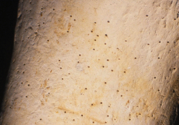
Figure 32: (l) Close-up photograph showing localised surface porosity on one of the upper limb bones recovered. This may have resulted from antemortem disease processes rather than taphonomic processes.
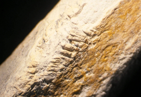
Figure 33: (r) Close-up photograph showing taphonomic effects on one of the tibia recovered.
Surface injury Localised injury caused by the teeth of Parrot fish from families Scuridae or Labridae was evident. Other markings consistent with unknown scavengers were visible. Extensive surface scratching most likely caused by abrasion of sand or reef debris could also be seen. Various bones showed surface markings of unknown origin.
Surface deposition Surface examination of the bones revealed encrustation with Bryozoa
and occasional Annelida.
Surface stains A green stain noted on a particular bone was analysed and confirmed to be from long-term contact with a copper alloy (e.g. brass).
Bone matrix infiltration Some material was invaded by a rock-boring sponge, resulting in the disintegration and destruction of bone. The medullary cavities of long bones were filled with coarse granular reef debris.
Exposure injury Cracking, flaking and shedding of surface bone material occurred in the laboratory as the bones dried out.
Descriptions of bone material being subjected to water environments have been published (Ascenzi and Silvestrini 1984), but these have little relevance to the Pandora remains. Skeletal trauma caused by marine scavengers is not well known (Iscan and McCabe 1995). The taphonomic changes present in the Pandora skeletal material examined here will be largely observational descriptions.
Examination of the Pandora skeletal remains demonstrated several taphonomic effects specific to the environment from which they came. Taphonomy was not one of the primary objectives for this study. The taphonomic effects of the skeletal material are largely descriptive.
Surface injury
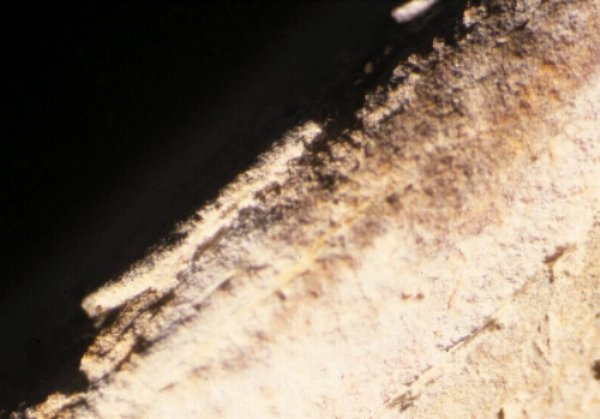
Some of the bones from the Pandora collection demonstrated significant physical change caused by long-term abrasive action. Such abrasion is the direct result of agitation between the bone itself and sand or reef debris. Essentially, this causes a smoothing of any jagged or sharp edges originally present on the surface of the bone, and the exposure of cancellous bone. As expected, the rate of abrasive destruction appears to be inversely proportional to the density of bone. This means that abrasive forces will effect thin trabecular or cancellous bone more rapidly than compact bone.
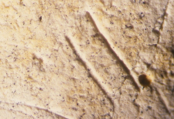
Localised surface injury was also evident from scavenger activity. Several types of markings were observed on some of the bones, probably caused by the teeth of fish, the exact species of which could not be determined. Numerous other postmortem markings of unknown origin were observed on the surface of the remains.
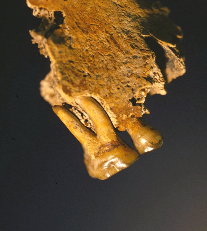
Figure 34: Close-up photograph showing repeated surface markings originating from unknown marine scavengers (left)
Figure 35: Close-up photograph showing parallel surface markings likely originating from the central incisors (or beak) of unknown marine scavengers (right).
Surface deposition
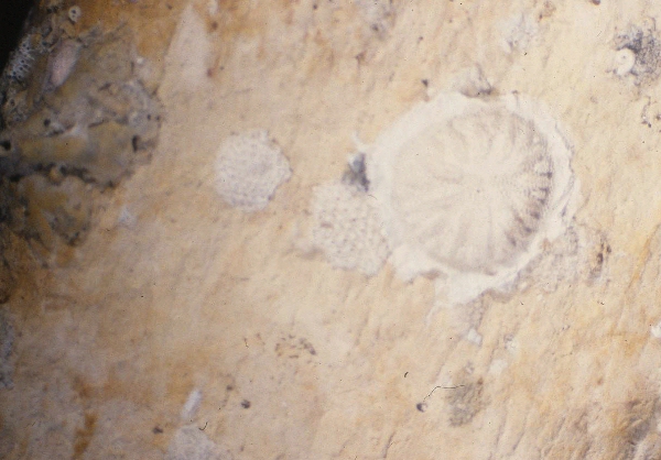
Some of the bones had occasional colonisations of Bryozoa and Annelida. Wood and Hodgson (1996) described similar encrustation on the 1986 material. Such marine life was consistently found on the highly degraded bones, and was accompanied by regions of orange-red staining. It is thought that this marine colonisation occurred in an aerobic environment. An aerobic environment would cause the oxidation of any iron objects known to be in close proximity to the bones, and may explain the coinciding orange-red stains.
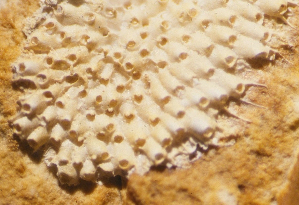
Figure 36: Close-up photograph showing the presence of marine colonisation (left)
Figure 37: Close-up photograph showing a bryozoan colony on one of the tibia recovered. Note the open opercula.
Surface staining
Surface staining was also evident on much of the material. The origin of the stains is unknown, although they were probably caused by oxidation of metallic alloys containing copper, zinc, iron and lead. Brass and iron objects were known to be on board the ship. The gunpowder room at the stern of the ship was also known to be lined with lead sheeting to avoid the potential propagation of sparks. Further studies could identify the origin of these stains by use of mass spectroscopy. Orange-red staining (discussed above) may indicate the bone was close to the surface of the sea-bed, in a more oxygen-rich environment. Objects lacking this type of staining were consistently better preserved and are thought to have been buried in the wreck in a deeper, more stable, anaerobic environment.
Some of the teeth belonging to the right maxillary fragment and mandible of Tom showed a yellow-brown staining. This is consistent with staining earlier described by Wood and Hodgson (1996) on the skeleton recovered in 1986, and is thought to have originated from long-term exposure to nicotine or tannin.
Figure 38: Lateral view of the right maxillary fragment belonging to Tom
Bone infiltration
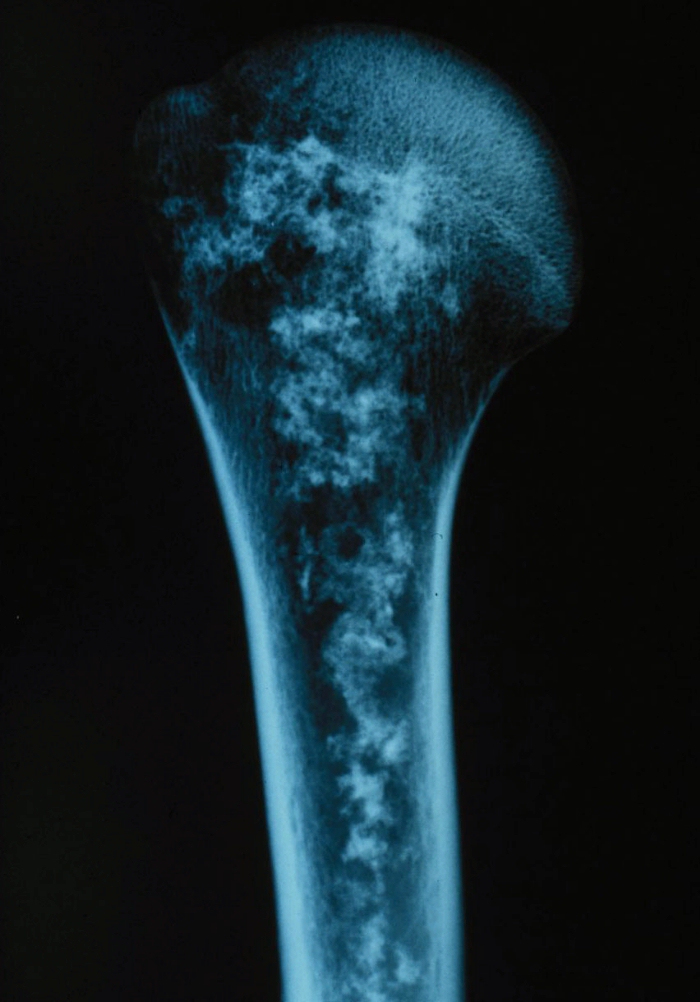
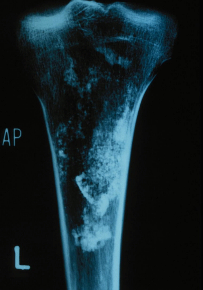
On occasion the medullary cavities of long bones were exposed due to excessive weathering of the proximal and/or distal ends. Where this was the case, medullary cavities were filled with compacted reef debris and sand. Upon drying the material, this coarse sand spilled out. Similar sand was found inside the cranium belonging to Harry. X-ray analysis demonstrated sand infiltration within other bones.
Figure 39: X-Ray of the proximal portion of a humerus. Note the infiltration of sand and reef debris.
Figure 40: X-Ray of the proximal portion of a tibia. Note the infiltration of sand and reef debris
Exposure injury
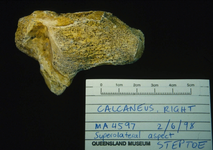
After being dried and exposed to the air, some bones experienced surface cracking and flaking after several months. This was mainly evident on the bones that were stained orange and highly degraded. The majority of the skeletal material did not degrade any further following conservation in the laboratory. If an iron cannon-ball did not undergo conservation within several days of exposure to air it would crumble into dust (Coleman pers. comm.). Conservation techniques utilised for the skeletal remains, and results obtained by the Queensland Museum were exceptional.
Figure 41 (l): Close-up photograph showing surface flaking and consequent taphonomic degradation of the mid-shaft surface of one of the ulna recovered
Figure 42 (r): Photograph of a right calcaneus. Note the degree of degradation [MA4597]
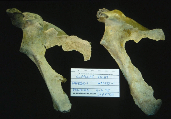
Figure 43 (l): Photograph showing two right scapulae [MA4868-1 and MA4531-1]. Note the degradation of the thin bone plate of the infra-spinous region.
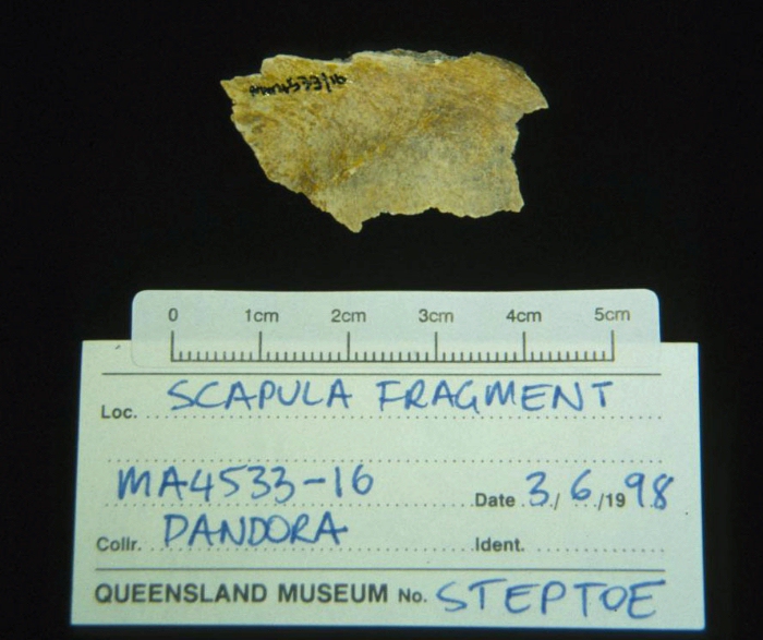
Figure 44 (r): Scapular bony fragment [MA4533-16].
Figure 45: X-Ray of tibia (center) and two fibula. Note the degradation of the proximal and distal portions of each.