PREVIOUS   NEXT   ISSUE   CONTENTS   HOME 

4.6 What is this? Unknown features observed

A variety of features unknown to us were observed in the tooth thin-sections. We would appreciate feedback from readers who may be able to identify what is seen in the following micrographs.

Figure 56   Figure 57
Figure 56: Tunnel-like features (white arrows) within the enamel of CDU-15 (cattle tooth). Could this be evidence of bioerosion in the enamel? The dentine is affected by intense bioerosion, going right up to the dentino-enamel junction whereas the enamel also exhibits extensive cracking. The tunnel-like features appear to emerge from the cracks. In Figure 56 the dark area does indicate cracks/tunnels filled with some opaque material. It is difficult to tell the features apart and further investigation would be needed to establish the nature of these. See also detail at higher magnification in Figure 57. (Image credit: H. Hollund)
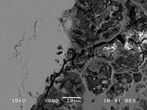
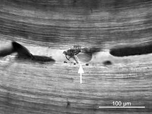
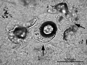
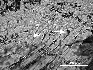
Figure 57: Tunnel-like features (white arrows) within the enamel of CDU-15 (cattle tooth). Could this be evidence of bioerosion in the enamel? See also Figure 56. (Image credit: H. Hollund)
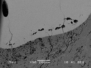
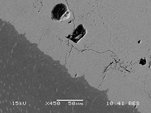
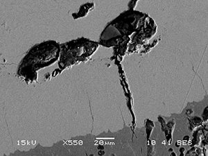
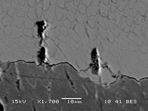
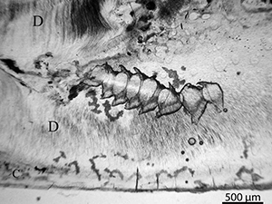
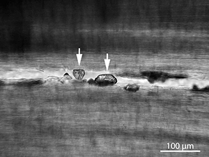
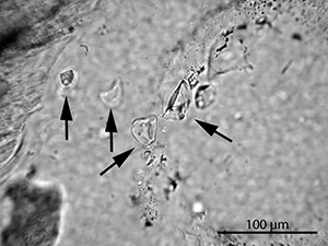
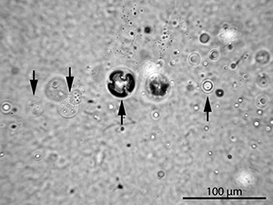
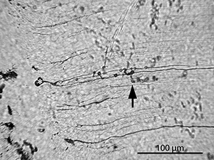
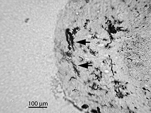
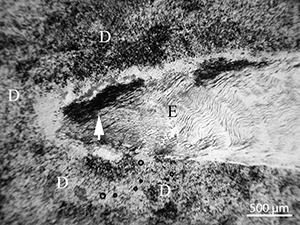
Figure 58   Figure 59 Figure 60   Figure 61 Figure 62
Figure 58: SEM-BSE images of globular cavities in the enamel of EIN-22. These are roughly 10-30 micron large, some empty, some containing inclusions. The cavities are located relatively close to the dentino-enamel junction and are sometimes associated with cracks. In several places the dentine is heavily bioeroded up to the dentino-enamel junction, where smaller cavities can be seen just on the border (Figure 61). See also Figures 59, 60 and 61. (Image credit: H. Hollund)
Figure 59: SEM-BSE images of globular cavities in the enamel of EIN-22. These are roughly 10-30 micron large, some empty, some containing inclusions. The cavities are located relatively close to the dentino-enamel junction and are sometimes associated with cracks. In several places the dentine is heavily bioeroded up to the dentino-enamel junction, where smaller cavities can be seen just on the border (Figure 61). See also Figures 58, 60 and 61. (Image credit: H. Hollund)
Figure 60: SEM-BSE images of globular cavities in the enamel of EIN-22. These are roughly 10-30 micron large, some empty, some containing inclusions. The cavities are located relatively close to the dentino-enamel junction and are sometimes associated with cracks. In several places the dentine is heavily bioeroded up to the dentino-enamel junction, where smaller cavities can be seen just on the border (Figure 61). See also Figures 58, 59 and 61. (Image credit: H. Hollund)
Figure 61: SEM-BSE images of globular cavities in the enamel of EIN-22. These are roughly 10-30 micron large, some empty, some containing inclusions. The cavities are located relatively close to the dentino-enamel junction and are sometimes associated with cracks. In several places the dentine is heavily bioeroded up to the dentino-enamel junction. Here, smaller cavities can be seen just on the border. See also Figures 58, 59 and 60. (Image credit: H. Hollund)
Figure 62: SEM-BSE images of globular cavities in the enamel of EIN-22. These are roughly 10-30 micron large, some empty, some containing inclusions, relatively close to the dentino-enamel junction and sometimes associated with cracks. (Image credit: H. Hollund)

Figure 63
Figure 63: Micrograph showing the remains of an insect within human tooth CAS-08. The insect must have been located in the pulp cavity, but as a result of how the tooth was cut it appears here as superimposed on the dentine. Our knowledge does not allow us to identify species and we also do not know how it got there; during decomposition, or perhaps post-excavation, in storage. Identifying such remains may give important information on taphonomy, season of burial etc.

Figure 64   Figure 65
Figure 64: Micrograph showing unknown inclusions in a crack in CAS-08. Considering that this tooth also contained the remains of an insect (Figure 63), could these be insect eggs? See also Figure 65. (Image credit: H. Hollund)
Figure 65: Micrograph showing unknown inclusions in a crack in CAS-08. Considering that this tooth also contained the remains of an insect (Figure 63), could this be an insect egg? See also Figure 64. (Image credit: H. Hollund)

Figure 66   Figure 67   Figure 68
Figure 66: Micrograph of inclusions within the pulp cavity of EIN-23. These inclusions represent a variety of sizes and shapes. Most are probably fungal spores, spore sacks and yeast cells. The possibility that some of the circular features are air bubbles cannot be excluded. See also Figures 67 and 68. (Image credit: H. Hollund)
Figure 67: Micrograph of inclusions within the pulp cavity of EIN-23. These inclusions represent a variety of sizes and shapes. Most are probably fungal spores, spore sacks and yeast cells. The possibility that some of the circular features are air bubbles cannot be excluded. The dark circle in the middle of the image is an air bubble. See also Figures 66 and 68. (Image credit: H. Hollund)
Figure 68: Micrograph of inclusions within the pulp cavity of EIN-23. These inclusions represent a variety of sizes and shapes. Most are probably fungal spores, spore sacks and yeast cells. The possibility that some of the circular features are air bubbles cannot be excluded. The dark circle in the middle of the image is an air bubble. See also Figures 66 and 67. (Image credit: H. Hollund)

Figure 69
Figure 69: Micrograph of sample EIN-10 showing cuffed circular shapes located along the length of two dentinal tubuli. Some of the circles appear directly on the tubuli, others at the end of what looks like enlarged lateral branches. In addition a possible microbial tunnel, similar to what has been observed in many of the teeth, is emerging from one of the tubuli (arrow). The globular shape at the end of the tubuli where it meets the layer of Tomes and the cemento-dentinal junction, may also be an MFD (asterisk)

Figure 70
Figure 70: This micrograph of an area of cementum in EIN-23 shows cementocyte lacunae that seem bloated and enlarged, with tree-like structures and fine branches emerging from them. Could these be MFD caused by microbes?