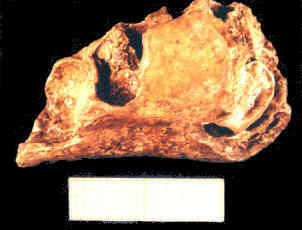
Plate 25: Sima de las Palomas: fragment of a child's mandible CG-7 Scale = 2cms (Photo M.J.Walker)
Hominid fossils from Sima de las Palomas are more numerous (Table 7). Several have been excavated systematically in the uppermost part of the breccia column, a few metres above where CG-1 (Plate 9, Plate 10) was plucked out by a spelaeologist in 1991; some show signs of burning (e.g. Plate 23 and Plate 24), others like CG-1 do not. Many were found during clearing or sieving dirt and rubble strewn liberally by miners on the hillside or in the mine level and main chamber. Archaic "pre"-Neanderthal bones were found in dirt and rubble in the main chamber and mine level. Many of these very light bones had undergone great heat; they are pale bluish-grey and give out a metallic "ring" (like porcelain) when tapped, and burnt teeth have crazed or shattered enamel (Blackish stains due to manganese oxide precipitated from percolating water are distinctively different).
As well as items in Table 7, other fragments of long-bone shafts may well be human, despite lacking distinctively human anatomical features, because great cortical thickness and narrow medullary cavities conform to a Middle-Upper Pleistocene hominid pattern that is uncommon in other mammals of human size.
Cranial skeletal morphology indicates presence of both Neanderthal and archaic, "pre"-Neanderthal hominid forms, though many teeth, postcranial, and even cranial bones could belong to either. Mandibular remains demonstrate presence of a Neanderthal adult (CG-1) and baby/foetus, both from the upper breccia, and two pre-Neanderthal adults (CG-6, male; CG-2, female) and a child 9-10 years old (CG-7) from unstratified mine rubble. Of these five, CG-6 and CG-2 show signs of burning. Teeth and an arm bone from the upper breccia, and an axis vertebra, indicate presence of at least one Neanderthal juvenile which could be the same as the CG-7 jaw fragment or perhaps represent a slightly older child or young adolescent.
CG-1 occurred 4-5m below the top of the breccia column. Just below CG-1, a uranium-thorium age of 50-60,000 years was estimated from an aragonite crystal. The fragmentary CG-1 mandible was cemented in breccia to both maxillary dental arcades (Plate 9, Plate 10); almost all permanent teeth seem present though few measurements can be taken as yet. An elastomere cast was made of the fossil in its original state. Cleaning and separation of its fragments are underway to restore them to their proper anatomical positions. Alveolar processes survive of maxillary palatal and incisive regions. The mandible is represented by a fragmentary left body, and more complete right body and ascending ramus despite fractures and deformation, doubtless caused by post-mortem sedimentary pressure. The right mandibular canine lies laterally, as in Neanderthals, at the angle of inflexion between frontal and lateral portions of the dental arcade. The mandible lacks mental eminences; its mental symphysis recedes posteriorly. Another typically Neanderthal feature is the posterior situation on the mandibular body of the mental foramen (below the posterior premolar-first molar separation). As in Neanderthals, the canine tooth lies directly above the limit of a fossa on the mandibular lower border for insertion of the anterior belly of the digastric muscle (both muscle and fossa are larger in modern people). As in Neanderthals, a retromolar gap separates the wisdom tooth from where the myelohyoid line and temporal crest unite on the ascending ramus. Cleaning the medial surface of the mandibular ramus shows too much damage has taken place for preservation of the mandibular foramen and, therefore, for its shape to be known (commonly oval-horizontal in Neanderthals, Stringer et al. 1984). Front teeth show heavy attrition, as is common in adult Neanderthals, and all the teeth seem large, though as yet it has only been possible to measure mesiodistal dimensions of the right mandibular medial incisor (5.1mm), left maxillary canine (8.5mm) and right mandibular canine (8.8mm). This last high value is almost without equal, apart from one Krapina tooth, in mandibular canines published from the Upper Pleistocene and early Holocene of Europe and western Asia. All three values have comparable Middle and Upper Pleistocene ones (cf. de Lumley-Woodyear 1973; Frayer 1978) and exceed Holocene values (cf. Ash 1987; Frayer 1978; Kieser 1990; Kraus et al. 1972; Olivier 1969). Cleaning demonstrates molar crowns are worn flat with dentine showing, suggesting a biological age of >30 years.
A small left symphyseal mandibular fragment, with empty anterior alveoli, of a very young infant (presumably Neanderthal) was excavated in layer 2a in the upper cutting extension. Maybe it belonged to the same very young infant as a left maxillary fragment found in mine rubble on the hillside, which measures 22.5mm anteroposteriorly and contains roots of the canine and both milk molars (the posterior root of 1m is missing), and an empty incisor alveolus. Two small fragments of a slightly larger mandible were also excavated from layer Ia of the upper cutting extension and belong to a probably somewhat older child; cleaning of considerable adherent breccia is underway. Burning is not seen on the aforementioned fragments, nor on a left anterior mandibular fragment (37mm long) of a child's mandible (CG-7) found when clearing out rubble in the mine level (Plate 25).
Plate 25: Sima de las Palomas: fragment of a child's mandible CG-7 Scale = 2cms (Photo M.J.Walker)
The fragment fails to reach the mental symphysis and an unerupted adult canine crown at the break has a degree of calcification similar to that in modern 9-10 year-old children (according to X-rays taken in Murcia by Drs José María Pastor and Enrique Fernández whose assistance is greatly appreciated). The submaxillary fossa almost reaches this anterior break. Eversion of the lower mandibular border recalls CG-2 and CG-6 (see below). A double mental foramen lies beneath an empty alveolus for the posterior deciduous molar, where the height of the body is 23mm.
 Plate 26: Sima de las Palomas: mandibular fragment CG-6. Scale in cm (Photo M.J.Walker)
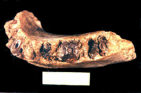 Plate 27: Sima de las Palomas: mandibular fragment CG-6. Scale = 2cm (Photo M.J.Walker)
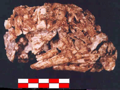
CG-6 is a severely burnt fragment of the left body of an adult mandible (Plate 26, Plate 27), found during clearance of dirt and scree piled up at the foot of the breccia column. Its 18mm thickness at 3M/1M far exceeds that of CG-1, and CG-6 is also much lower with a height of only 27mm at 3M/2M. The mental symphysis is vertical anteriorly, having a D-like cross-section 22mm high and 16mm thick. The digastric fossa is well-marked and the submaxillary fossa is triangular. The mental foramen's large size (8x5mm) indicates amplification by great heat. All 3P-3M tooth roots are preserved. Empty anterior alveoli correspond to a canine and lateral and medial incisors lost after death. Clearly, their presence in life excludes any possibility that anterior symphyseal verticality reflects only bony remodelling following anterior alveolar resorption; this was one conjecture to interpret symphyseal verticality in KRM-41815 from Klasies River Mouth rock-shelter 1b in South Africa, though CG-6 is too robust anyhow for remodelling to sculpt a "pseudo-chin". The symphyseal development of CG-6 may be comparable to exceptional mandibles from Atapuerca Sima de los Huesos (e.g. AT-300) or Caune d'Arago. Alveolar margin erosion and resorption occurred near premolar and molar necks; no crowns are preserved. A (taurodont) anterior premolar has a wide pulp cavity in its single root. CG-6's canine lies on the inflexion between frontal and lateral parts of its tooth row, which is a primitive pre-Neanderthal condition, in sharp contrast to CG-1's typically Neanderthal, lateral, situation, behind the angle of inflexion between frontal and lateral regions of the tooth row.
Mandibular body height (Table 8) at the second molar (HBM2 = Rudolf Martin's measurement "69(2)": Martin 1928, 663) is 30.32mm for CG-1 and 27.04mm in CG-6. Body heights at the wisdom tooth (HBM3) are 30.54mm and 24.08mm, between the second and third molars (HBM2M3) 29.64mm and 26.82mm, and at the anterior premolar (HBP3) 30.62mm and 28.01mm. These, and those for CG-1 and CG-2, may be compared with similar, if scantily published, values for other fossil mandibles (multivariate statistical comparisons are underway but results are not yet available). Broadly comparable are Atapuerca Sima de los Huesos (M3 body heights for AT-1,-75,-250,-300,-505,-605,-607,-792,-950,-888 are 28.9, 28.7, 29.5, 30.1, 25.6, 32.9, 27.2, 30.5, 28.2, 35.9mm: Rosas 1997) or Caune d'Arago 2 in French Catalonia (27.2mm high at the lateral eminence).
CG-2 is a burnt adult right mandibular body fragment, found when clearing mine rubble off the hillside. Parts of most of the teeth remain but only the middle molar crown is intact enough for inspection of occlusal attrition. The lateral incisor has a high, archaic, buccolingual dimension which at 8.0mm surpasses that of CG-1. The symphysis is vertical, as in CG-6, with no mental eminence (bony resorption cannot explain verticality here any more than in CG-6); perhaps Banyolas in Catalonia is comparable (nowadays dated to 45-50,000 years ago). Below the first molar's posterior edge there is a double mental foramen; here the body is only 14mm thick albeit 28mm high. However, eversion of its lower border below the posterior teeth resembles that of CG-6 and its height of 27mm between the two rear molars is similar, though even here the body is only 14.6mm thick. CG-2 has a smoother surface with fewer muscle markings than CG-6, and its dental arcade at 51mm is shorter than CG-6's 57mm. Very likely CG-2 is female and CG-6 male. Their differences recall Atapuerca mandibular variation (cf. Rosas 1997). CG-2 is adult because the wisdom tooth had erupted; this shows a wide pulp cavity. The second molar has dentine exposed by attrition over one-third of the occlusal surface. Although front tooth crowns are missing, there are neither signs of anterior tooth loss before death, nor of mandibular body atrophy with loss of height, any of which might have been expected in a Neanderthal so elderly that even its back teeth had become worn down after attrition of its front teeth. Alveolar margin erosion and partial resorption occur between the second and third molars and traces of erosion exist from the rear premolar to the medial incisor. Table 9 gives CG-2's odontometric measurements.
| CG-2 teeth | I1 | I2 | C | P3 | P4 | M1 | M2 | M3 |
|---|---|---|---|---|---|---|---|---|
| mesiodistal crown dim. in mm | 5.0 | 5.8 | 7.0 | 5.5 | 6.5 | 11.0 | 11.0 | 11.0 |
| buccolingual crown dim. in mm | - | - | - | - | - | - | (9.5 ?11*) | 11.0 |
| crown height in mm | - | - | - | - | - | - | - | 5.0 |
| mesiodistal dim.at neck in mm | 4.0 | 5.0 | 6.0 | 5.0 | 5.0 | 9.0 | - | - |
| buccolingual dim. at neckin mm | 7.0 | 8.0 | 9.0 | 8.0 | 8.0 | 9.0 | - | - |
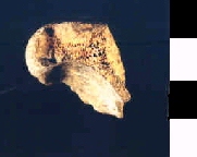
Two small burnt frontal fragments show supraorbital tori, found while clearing the sloping scree pile at the foot of the breccia column (CG-15), and mine rubble from the hillside (CG-14). CG-14 is the central part of a left torus, 11.0mm thick, extending from the lateral wall of a medial notch - starting beyond the medial point of the orbital margin and ending before reaching its midpoint. At those points, Steinheim, most Atapuerca Sima de los Huesos and Neanderthal tori are 10-17mm thick (Arsuaga et al. 1997). CG-14's flat superior surface measures 18.5mm anteroposteriorly. CG-15 (Plate 24), slightly larger, shows parts of the right orbital roof and right anterior cerebral fossa floor, being 36mm long anteroposteriorly and 32mm mediolaterally in the anatomical position. Of the damaged 29mm of its superior orbital margin, only the lateral 12mm retain part of a prominent supraorbital torus that continues into a frontal trigone receiving a superior temporal line. Its shape resembles Steinheim, though its 7.7mm thickness is around 2mm less than Steinheim's. Perhaps CG-14 is male and CG-15 female. Found also on the hillside is a burnt fragment (34x22x4.5mm) of frontal bone behind the left lateral frontal protuberance, showing an impression of the anterior branch of an ascending middle meningeal artery and a tiny part of a coronal suture.
A burnt left temporal squame and sigmoid fossa (CG-12; Plate 23), found during clearance of the scree slope at the foot of the breccia column, belonged to an adolescent or young adult whose temporoparietal and occipitomastoid sutures were unfused. Its outer surface has strongly marked ridges for insertion of temporalis and an elongated prominent mastoid crest. Its greatest height is 48mm and its anteroposterior length 62mm. In outline it arches upwards as in Neanderthal and Atapuerca Sima de los Huesos temporals. The articular eminence is not prominent, the zygomatic tubercle reaching 15mm below the upper surface of the root of the zygomatic process and 8mm below the roof of the sigmoid fossa. A minute tympanic fragment adheres to the posterior surface of the tuberosity but is broken posteriorly. The sigmoid fossa is 22mm wide and 19mm long. The zygomatic process is broken near its root and the mastoid process and petrous temporal parts are missing. (A loose, but unburnt, mastoid process was found on the hillside.) A nutrient foramen is present on the zygomatic process just above the anterior zygomatic tubercle, and another enters the temporal squame between the insertional ridges for temporalis and the anterior border of the root of the zygomatic process. Behind the latter, a hole (2.5mm wide and 8mm long) runs through the entire bone just in front of the external auditory meatus; it was probably caused after death because the internal squamous surface shows many pores, probably diagenetic in origin. Flattish articular eminences may be a Neanderthal apomorphy with Bilzingleben Castel di Guido, Petralona, Steinheim and Atapuerca Sima de los Huesos antecedents (Martínez and Arsuaga 1997).
Clearing mine rubble on the hillside in 1997 brought to light a large left maxillary fragment with a broad lower nasal margin as in Neanderthals but missing its teeth (it is under investigation and awaiting an identification number). Among thick, burnt, cranial fragments from rubble on the hillside are a burnt rectangular Wormian bone, CG-10, with four unfused sutures (24x20x7mm); a left parietal fragment, CG-5, (39x34x7.5mm) with vestiges of both sagittal and lamboid sutures, a nutrient foramen, and an impression of the posterior part of a middle meningeal artery; part of a right parietal, CG-4, (57x47x5.5mm) showing the parietotemporal suture and impressions of posterior branches of the middle meningeal artery; and a pentagonal fragment of occipital squame, CG-3, (47x 47x6mm) with the internal occipital protuberance (7mm thick) and part of the right lambdoid suture; as well as other probable fragments, including part of a zygomatic and greater wing of sphenoid.
Loose teeth are extremely important because odontometric and morphological characteristics define several as undoubtedly Neanderthal - some of these were excavated in the uppermost part of the breccia column, others occurred among mine rubble. Many odontometric measurements show teeth from our upper cutting have Neanderthal values (surpassing modern ones). Odontometric measurements were made by several of us because sometimes slight differences in methodology affect results. We briefly present permanent incisors, permanent canines, premolars, permanent molars, deciduous incisors, deciduous canines, and deciduous molars.
© Internet Archaeology
URL: http://intarch.ac.uk/journal/issue5/walker/4.2.html
Last updated: Wed Dec 23 1998