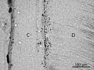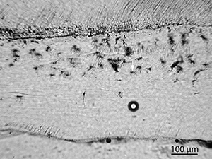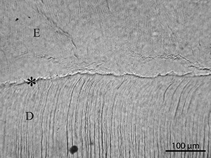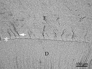
The following sections describe the observations made concerning both biological and non-biological diagenetic features, illustrated by micrographs. The histological indexes are reported in Table 2. In addition, results from SEM-analyses in the form of backscatter electron images (SEM-BSE) and elemental spectra (SEM-EDX) are presented.

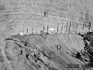
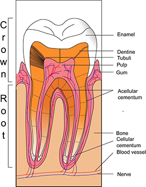
Before assessing diagnetic alterations, it is necessary to be familiar with the main micro-anatomical features of teeth as seen in histological thin-sections. Teeth consist of three different types of tissues: enamel, dentine and cementum, as seen in the schematic drawing in Figure 2. The bulk of the element is made up of dentine. The roots are covered with cementum and the crown is capped by enamel. Dentine is avascular and acellular but the tissue is perforated by long odontoblast processes connected to cells within the pulp cavity. These tubuli are similar in diameter to bone canaliculi and connect the dentine to the vascular system. The cementum is partially cellular, containing cementum-forming cells called cementocytes. The enamel is almost entirely inorganic and is a denser and more crystalline material than the other tissues (Hillson 1986). Figures 3 to 7 show micrographs that illustrate micro-anatomical features of teeth as seen in longitudinal cross-sections of whole teeth.
