PREVIOUS   NEXT   ISSUE   CONTENTS   HOME 

4.2 Bioerosion

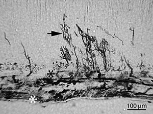
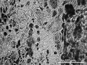
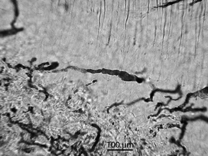
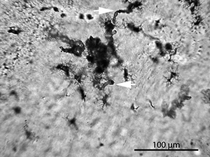
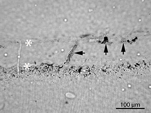
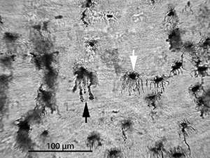
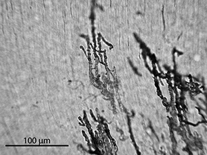
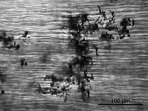
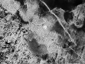
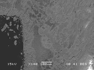
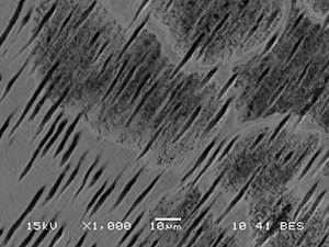
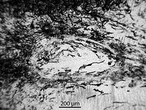
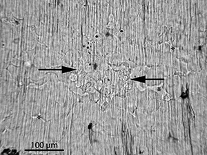
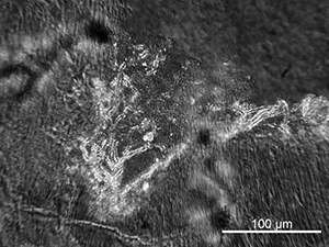
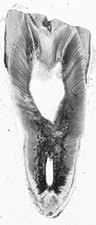
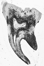
In the transversal thin sections of the Eindhoven bone samples, microbial tunnelling was observed as Hackett's budded and linear longitudinal tunnels. The tunnels in the dentine and cementum appear largely similar, with a similar morphology and size to those in bone. Several of the teeth displayed possible analogues to the Wedl type 2 tunnels found in bone (Trueman and Martill 2002) in the form of enlarged canaliculi within the cementum. Within the dentine, destruction also appeared in the form of enlarged dentinal tubuli.

Figure 8   Figure 9
Figure 8: Scanned thin-section of sample EIN-15 showing the pattern of microbial tunnelling. The tunnels (dark areas) are mainly found within the cementum, the apex (root tip) and along the root canal
Figure 9: Scanned thin-section of sample EIN-22. Here, the attack is more extensive than in EIN-15 (Figure 8) and the microbes are also entering from the surface of the pulp cavity penetrating the whole way to the dentino-enamel junction

Figure 10
Figure 10: Micrograph of sample EIN-23. The cementum is completely bioeroded, mainly in the form of globular MFD. Despite the cemento-dentinal junction (black asterisk), the bacteria are penetrating into the dentine. The micro-organisms seem to enter the tooth at the cementum surface (white asterisk). Tunnelling into the dentine is in the form of enlarged dentinal tubuli (arrow) with branches. See also detail in Figure 16

Figure 11
Figure 11: Micrograph of sample EIN-05 showing globular-shaped tunnels within the dentine. These are of the same size and shape as the linear longitudinal tunnels (approximately 10 µm) and budded tunnels (up to 100s of µm) seen in bone. In the middle of the image a pristine area of dentine can be seen, with the dentinal tubuli clearly visible

Figure 12
Figure 12: Micrograph of sample EIN-16. Destructive foci are observed as several connected oblong shapes

Figure 13
Figure 13: Micrograph of sample EIN-23. Elongated destructive foci (arrows) stretch out from cementocyte lacunae and cancaliculi

Figure 14
Figure 14: Micrograph of sample EIN-23. Three destructive foci within the cementum where the micro-organisms seem to have entered from the cementum surface. The white asterisks mark the cementum surface and the cemento-dentinal junction

Figure 15
Figure 15: Micrograph of sample EIN-04. What can be described as Wedl type 2 tunnels are found within the cementum. These appear as enlarged canaliculi, first described in bone by Trueman and Martill (2002). However, these may also be caused by acid etching, as suggested by Fernández-Jalvo et al. (2010) for bone samples. This may also be the case here (black arrow). Alternatively, the canaliculi may appear enlarged whereas this is caused by staining along the canaliculi. The fine central line may be the original canaliculi and the broader darker band being caused by infiltrations. The white arrow indicates an unaffected cementocyte lacuna

Figure 16
Figure 16: Micrograph of sample EIN-23. Destructive foci in the dentine appear as enlarged dentinal tubuli and lateral branches

Figure 17
Figure 17: Micrograph of sample EIN-23. Destructive foci in the form of enlarged lateral branches of the dentinal tubuli

Figure 18
Figure 18: Micrograph of sample EIN-22. An area with large, globular MFD with a grainy interior can be seen in the middle of the image (asterisk). The pattern of the dentine microstructure is visible within the foci, in a way similar to lamellar tunnels in bone (Hackett 1981) and what was observed by Bell et al. (1991) in dentine. See also SEM-BSE images in Figures 19 and 20

Figure 19   Figure 20
Figures 19: SEM-BSE image of the large MFD seen in Figure 18. The SEM-BSE images show that the destructions consist of a demineralised area with a border of more highly mineralised material. See SEM-BSE image in Figure 20 for higher magnification. (Image credit: H. Hollund)
Figure 20: SEM-BSE image of the large MFD seen in Figure 18 and 19. At this level of magnification it is possible to see that the interior of the MFD contains numerous fine pores (roughly 1 micron across), as found within bone MFD by Jackes et al. (2001), believed to represent bacterial tunnels. (Image credit: H. Hollund)

Figure 21
Figure 21: Micrograph of sample EIN-06. As pathologies will change the physico-chemical and/or microstructural characteristics of teeth, these may also affect the pattern of microbial attack. Here, a circular deformation is visible within the dentine; a pulp stone. These may arise as an age change or accompany inflammatory or degenerative changes in the pulp. Calcification of the pulp results in the formation of discrete, approximately circular mineralised masses which may later be incorporated into the dentine (Marsland and Browne 1975). The microbial tunnelling can be seen to follow the circular lamellar microstructure of the imbedded pulp stone

Figure 22
Figure 22: Micrograph of sample EIN-15. This sample displays another commonly occurring pathology, a developmental defect leading to zones of interglobular and globular dentine. These are areas of unmineralised or hypomineralised dentine where globular zones of mineralisation have failed to fuse into a homogeneous mass within mature dentine. The condition is especially prevalent in human teeth as a result of vitamin D deficiency or exposure to high levels of fluoride at the time of dentine formation (Nanci 2003). In a few samples, microbes seem to have targeted the less mineralised inter-globular dentine, as can be seen in this micrograph. The two arrows mark a cluster of MFD located in an area with inter-globular dentine. Inter-globular areas are indicated with an asterisk. These MFD are located in an area of the crown dentine, directly beneath an enamel lesion. This tooth displays otherwise dense bioerosion around the root canal and in the cementum, while there are few MFD above the pulp cavity. This may suggest that the MFD in this area were caused by micro-organisms that entered via the lesion

Figure 23
Figure 23: Micrograph of sample CDU-15, viewed in polarized light. This is a Neolithic cattle tooth, the only sample to display clearly identifiable fungal tunnelling. Round, transparent fungal structures can be seen, as well as hyphae/tunnels penetrating into the dentine from the pulp cavity surface, appearing as the characteristic Wedl type tunnels (Hackett 1981; Marchiafava et al. 1974; Jans 2005). The hyphae/tunnels are clearly visible in polarized light as they are lined with a birefringent material, possibly calcite as this is a known precipitate on and in soil fungal structures (Burford et al. 2006). See also Figures 40, 41, 42 and 43.