PREVIOUS   NEXT   ISSUE   CONTENTS   HOME 

4.4 Generalised destruction and cracking

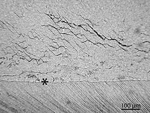
Since dentine and cementum are chemically similar materials to bone, these are also prone to mineral dissolution and low pH. This was seen in 12 samples (35%) and is indicated by low GHI and high OHI, since this former index records both bioerosion and generalised destruction. Microcracking was not observed in dentine/cementum of any of the teeth, but large cracks are found, often in conjunction with generalised destruction.

Figure 45
Figure 45: Scanned thin-section of EIN-17 illustrating the effects of an extreme corrosive environment that has caused generalised destruction and complete loss of a large part of the roots. Despite this, the dentine directly beneath the enamel crown remains well-preserved, testifying to the resilience of enamel and the efficient protection it provides. A caries lesion can also be seen in the enamel. This has caused some staining and reaction in the dentine

Figure 46
Figure 46: Micrograph of sample EIN-11 showing the effect of generalised destruction on the dentinal microstructure. The striated structure of the dentine has 'faded' in the upper half of the image. In the lower half, the tubuli and their lateral branches, appear stained and possibly enlarged. It is not possible to determine whether or not this is due to acid etching alone, or bioerosion prior to the etching

Figure 47
Figure 47: Micrograph of sample EIN-03. An area of generalised destruction, including staining and large cracks is shown. The cementum is lost in some areas and is detaching at the cemento-dentine junction (arrow). Sample preparation may also have caused part of this. The asterisk marks the root canal

Figure 48
Figure 48: Micrograph of EIN-08. Micro-fissures are present in the enamel. The enamel of the teeth studied was frequently cracked to different degrees