PREVIOUS   NEXT   ISSUE   CONTENTS   HOME 

4.3 Inclusions and infiltrations

Several of the teeth contained orange and opaque irregularly shaped inclusions within vascular canals, osteocytes, canaliculi and dentinal tubules. These were found by chemical analysis (SEM-EDX) of selected Eindhoven samples to contain iron and manganese (see Figure 32). Birefringent material, most likely calcite, was also observed filling microbial tunnels and pores in a couple of teeth. In addition, possible calcite-containing inclusions were found within the pulp cavity. Inclusions of framboidal pyrite (FeS2) were observed in one tooth (Figures 24-26). Both bones and teeth from Eindhoven were affected by orange and brown staining to varying degrees. This may be due to infiltrations by metal compounds and/or humic factors. Chemical analysis of infiltrations observed in four teeth and three bone samples from the site detected iron and manganese (Figure 35). Most samples contained remains of various fungal structures within canals and cavities.

Figure 24   Figure 25
Figure 24: Micrograph of ZWO-01. This human tooth contained many framboidal pyrite (FeS2) grains on the surface of the pulp cavity and root canals, in places almost completely filling the root canals. The cementum and dentine of this tooth is stained yellow and orange indicating infiltration by iron compounds, possibly a result of partial oxidation of pyrite (see discussion in Hollund et al. 2012b). See also detail in Figure 25. (Image credit: H. Hollund)
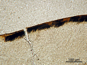
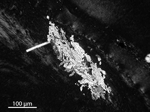
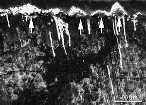
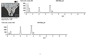
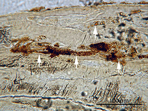
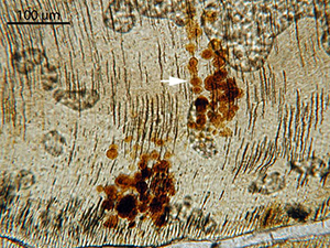
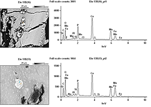
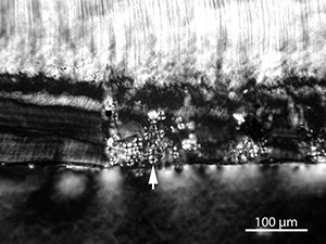
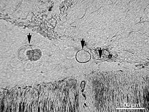
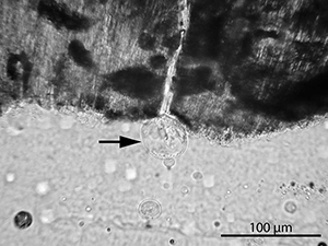
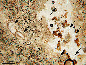
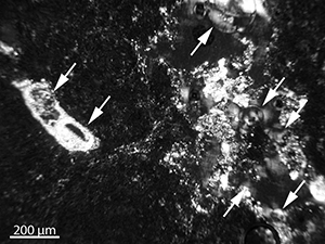
Figure 25: Micrograph of ZWO-01. Detail of the same tooth as shown in Figure 24. This human tooth contained many framboidal pyrite (FeS2) grains on the surface of the pulp cavity and root canals, in places almost completely filling the root canals. The cementum and dentine of this tooth is stained yellow and orange indicating infiltration by iron compounds, possibly a result of partial oxidation of pyrite (see discussion in Hollund et al. 2012b). (Image credit: H. Hollund)

Figure 26
Figure 26: Micrograph showing an area of the enamel surface of ZWO-01 which is stained black and orange, possibly infiltrated by pyrite and iron oxides (see also Figures 24 and 25)

Figure 27
Figure 27: Micrograph of sample EIN-10 in polarized light. Empty microbial tunnels are filled with a birefringent material. Analysis by SEM-EDX detected calcium, carbon and oxygen (Figure 29); most likely calcite (see also Figure 28)

Figure 28
Figure 28: SEM-BSE image of sample EIN-10. Empty MFD are filled by dense material, probably calcite, as suggested by the birefringence in polarized light (see Figure 27) and SEM-EDX analysis (Figure 29)

Figure 29
Figure 29: SEM-EDX results; chemical spot analysis of inclusions in empty MFD, sample EIN-10 (see Figures 26-28)

Figure 30   Figure 31
Figure 30: Micrograph of sample EIN-13 in normal light. Small (approximately 14 μm) orange and translucent pyramid-shaped nodules are found, sitting on the surface of the pulp cavity and root canal. Some of the dentinal tubuli stretching out from the cavity are also filled with orange material. This material shows birefringence in polarized light (Figure 31) and was found by SEM-EDX to contain calcium, carbon, oxygen and sometimes manganese, iron and phosphorus (Figure 32). (Image credit: H. Hollund)
Figure 31: Micrograph of sample EIN-13 in polarized light, showing that some of the inclusive material seen in Figure 30 is birefringent. This material was found by SEM-EDX to contain calcium, carbon, oxygen and sometimes manganese, iron and phosphorus (Figure 32). (Image credit: H. Hollund)

Figure 32
Figure 32: SEM-EDX results; chemical spot analysis of inclusions in EIN-13 (see Figures 30 and 31)

Figure 33   Figure 34
Figure 33: Micrograph of sample EIN-13. The sample contains orange perfectly round inclusions (arrows) and roughly circular stains (see Figure 34). These were found by SEM-EDX to contain iron and manganese (Figure 35). The inclusions are found within empty MFD in the cementum and dentine, whereas the stains mainly seem to follow the length of the dentinal tubuli. (Image credit: H. Hollund)
Figure 34: Micrograph of sample EIN-13. The sample contains roughly circular stains in addition to the round inclusions seen in Figure 34. Both stains and inclusions were found by SEM-EDX to contain iron and manganese (Figure 35). The inclusions are found within empty MFD in the cementum and dentine, whereas the stains mainly seem to follow the length of the dentinal tubuli. (Image credit: H. Hollund)

Figure 35
Figure 35: SEM-EDX results, inclusions and stains in EIN-13 (see also micrographs, Figures 33 and 34)

Figure 36
Figure 36: Micrograph of sample EIN-09 in polarized light, showing starch-grains within a crack in the cementum surface. The grains display the starch-specific extinction cross in polarized light (Piperno et al. 2004)

Figure 37
Figure 37: Micrograph of sample EIN-08, with remains of fungal fruiting bodies and fungal hyphae (arrows) within the pulp cavity

Figure 38
Figure 38: Fungal fruiting body on pulp cavity surface of EIN-24. The hyphae is located in a crack in the dentine

Figure 39
Figure 39: Yeast cells within the pulp cavity of cattle tooth CDU-15. This double form is a yeast cell reproducing by a division process called budding

Figure 40
Figure 40: Micrograph of sample CDU-15 showing transparent, round fungal structures within the pulp cavity. Tunnels (white arrows) can be seen penetrating the dentine. As seen in Figure 23, the hyphae/tunnels are lined with birefringent material, probably calcite and are thus visible in polarized light (Figure 41)

Figure 41
Figure 41: Micrograph of sample CDU-15, same view as Figure 40 but in polarized light. The tunnels, which seem to be caused by the action of fungal hyphae, are lined with a birefringent material that is visible in polarized light (see also detail in Figure 23).

Figure 42   Figure 43
Figure 42: Micrograph of sample CDU-15, viewed in normal light. In normal light, fungal fruiting bodies (arrows) are seen within pores/cracks in the dentine, associated with masses of orange and translucent material. This material displays a bright white birefringence in polarized light. As with the fungal hyphae seen in Figure 41, one of the fungal fruiting bodies is lined with the material. No analyses were carried out but it is likely that this is calcite, with some compounds containing iron and/or manganese, as seen for sample EIN-13 (Figures 30, 31, 32) and EIN-10 (Figures 27, 28, 29). (Image credit: H. Hollund)
Figure 43: Micrograph of sample CDU-15, viewed in polarized light, showing that some of the fungal fruiting bodies (arrows) also seen in Figure 42, are lined with a bright white birefringent material. No analyses were carried out but it is likely that this is calcite, with some compounds containing iron and/or manganese, as seen for sample EIN-13 (Figures 30, 31, 32) and EIN-10 (Figures 27, 28, 29). (Image credit: H. Hollund)

Figure 44
Figure 44: Micrograph of sample CDU-15 showing fungal spores within the pulp cavity